Explore
 Validate
 Learn
Learn Western blot
 Immunoprecipitation
Antibody data
- Antibody Data
- Antigen structure
- References [0]
- Comments [0]
- Validations
- Western blot [1]
- Immunohistochemistry [2]
Submit
Validation data
Reference
Comment
Report error
- Product number
- AP21656SU-S - Provider product page

- Provider
- Acris Antibodies GmbH
- Proper citation
- Acris Antibodies GmbH Cat#AP21656SU-S, RRID:AB_10758596
- Product name
- anti Caspase-3 (Pro and Active)
- Antibody type
- Polyclonal
- Antigen
- Recombinant full-length human Caspase-3 protein (pro-form)
- Reactivity
- Human, Mouse, Rat, Canine
- Host
- Rabbit
- Vial size
- 50 µl
No comments: Submit comment
Supportive validation
- Submitted by
- Acris Antibodies GmbH (provider)
- Main image

- Experimental details
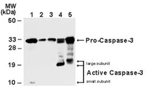
- Western blot analysis of Caspase-3. Lysates from Jurkat cells (lane 1), normal mammary tissue (lane 2) and surgical specimens from three invasive ductal carcinomas (lanes 3-5)were normalized for total protein content (50 µg/lane) and western blotted with anti-Caspase-3 (AP21656SU-S). The ~32 kDa pro-Caspase-3 protein was detected in all samples. Active/cleaved Caspase-3 was identified in Jurkat (10 kDa small subunit, lane 1) and two ductal carcinomas (14-21 kDa large subunit).
Supportive validation
- Submitted by
- Acris Antibodies GmbH (provider)
- Main image

- Experimental details
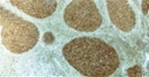
- Immunohistochemical analysis of Caspase-3 expression in formalin-fixed, paraffin-embedded human reactive lymph node using AP21656SU-S at 1/2000. Staining is seen in the apoptosis-prone germinal center B lymphocytes of follicles. In constrast little or no staining is seen in the surrounding long-live mantle zone lymphocytes.
- Submitted by
- Acris Antibodies GmbH (provider)
- Main image

- Experimental details
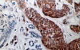
- Immunohistochemical analysis of Caspase-3 expression in formalin-fixed, paraffin-embedded human breast ductal carcinoma in situ using AP21656SU-S at 1/2000. Staining is seen in the the cancerous ducts, but not in the normal lobulus.