Explore
 Validate
 Learn
LearnPA3-009
antibody from Invitrogen Antibodies
Targeting: PDIA3
ERp57, ERp60, ERp61, GRP57, GRP58, HsT17083, P58, PI-PLC
 Western blot
 Immunocytochemistry
Antibody data
- Antibody Data
- Antigen structure
- References [3]
- Comments [0]
- Validations
- Immunocytochemistry [9]
- Immunohistochemistry [2]
- Other assay [6]
Submit
Validation data
Reference
Comment
Report error
- Product number
- PA3-009 - Provider product page

- Provider
- Invitrogen Antibodies
- Product name
- ERp57 Polyclonal Antibody
- Antibody type
- Polyclonal
- Antigen
- Synthetic peptide
- Description
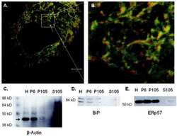
- PA3-009 detects ERp57 protein in human samples. PA3-009 has successfully been used in Western blot and immunofluorescence procedures. By Western blot, this antibody detects a 57 kDa protein representing ERp57 in rat liver homogenate. The PA3-009 immunogen is a synthetic peptide corresponding to residues I(490) Q E E K P K K K K K A Q E D L(505) of human ERp57. This peptide (Cat. # PEP-225) is available for use in neutralization and control experiments.
- Reactivity
- Human
- Host
- Rabbit
- Isotype
- IgG
- Vial size
- 100 μL
- Concentration
- Conc. Not Determined
- Storage
- -20°C, Avoid Freeze/Thaw Cycles
Submitted references ERp18 regulates activation of ATF6α during unfolded protein response.
Bromodomain inhibition exerts its therapeutic potential in malignant pleural mesothelioma by promoting immunogenic cell death and changing the tumor immune-environment.
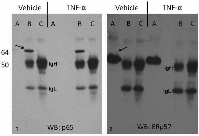
Tumor necrosis factor-α treatment of HepG2 cells mobilizes a cytoplasmic pool of ERp57/1,25D₃-MARRS to the nucleus.
Oka OB, van Lith M, Rudolf J, Tungkum W, Pringle MA, Bulleid NJ
The EMBO journal 2019 Aug 1;38(15):e100990
The EMBO journal 2019 Aug 1;38(15):e100990
Bromodomain inhibition exerts its therapeutic potential in malignant pleural mesothelioma by promoting immunogenic cell death and changing the tumor immune-environment.
Riganti C, Lingua MF, Salaroglio IC, Falcomatà C, Righi L, Morena D, Picca F, Oddo D, Kopecka J, Pradotto M, Libener R, Orecchia S, Bironzo P, Comunanza V, Bussolino F, Novello S, Scagliotti GV, Di Nicolantonio F, Taulli R
Oncoimmunology 2018;7(3):e1398874
Oncoimmunology 2018;7(3):e1398874
Tumor necrosis factor-α treatment of HepG2 cells mobilizes a cytoplasmic pool of ERp57/1,25D₃-MARRS to the nucleus.
Grindel BJ, Rohe B, Safford SE, Bennett JJ, Farach-Carson MC
Journal of cellular biochemistry 2011 Sep;112(9):2606-15
Journal of cellular biochemistry 2011 Sep;112(9):2606-15
No comments: Submit comment
Supportive validation
- Submitted by
- Invitrogen Antibodies (provider)
- Main image

- Experimental details
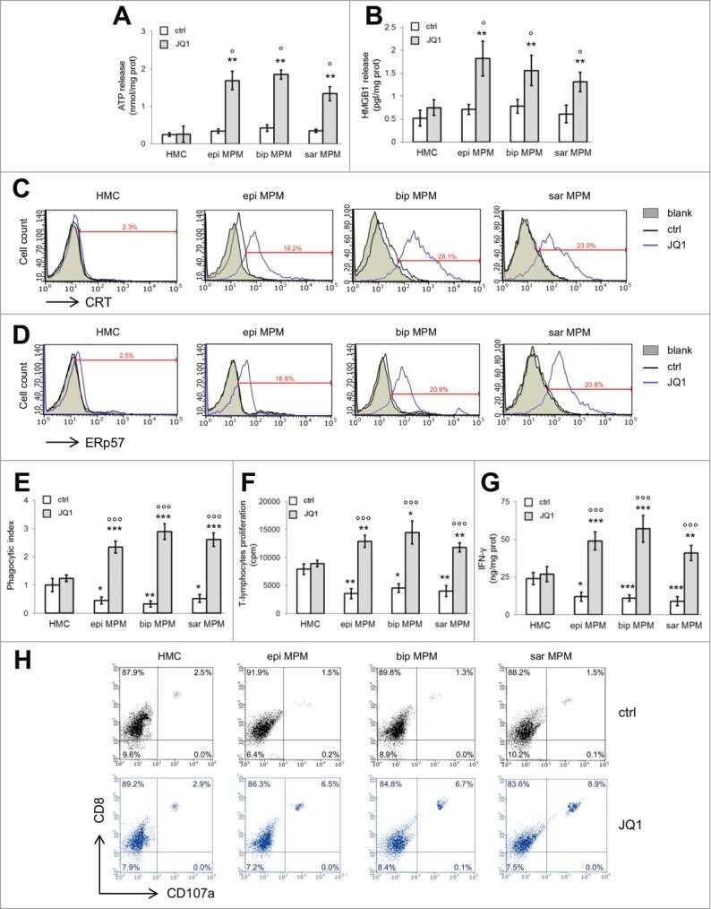
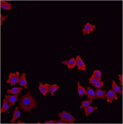
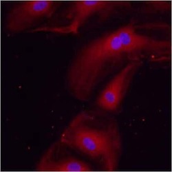
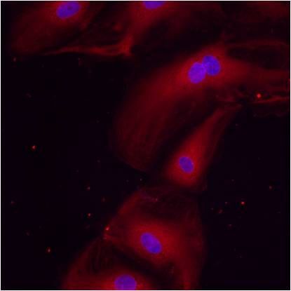
- Immunofluorescent analysis of ERp57 using anti-ERp57 polyclonal antibody (Product # PA3-009) shows staining in HMVEC Cells.

- Submitted by
- Invitrogen Antibodies (provider)
- Main image

- Experimental details
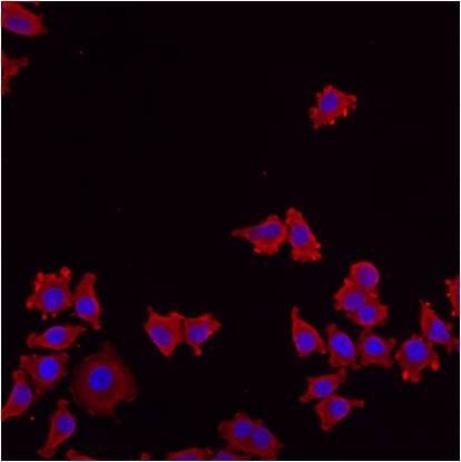
- Immunofluorescent analysis of ERp57 using anti-ERp57 polyclonal antibody (Product # PA3-009) shows staining in NS-1 Cells.
- Submitted by
- Invitrogen Antibodies (provider)
- Main image

- Experimental details
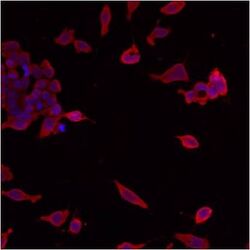
- Immunofluorescent analysis of ERp57 using anti-ERp57 polyclonal antibody (Product # PA3-009) shows staining in p19 Cells.
- Submitted by
- Invitrogen Antibodies (provider)
- Main image
- Experimental details
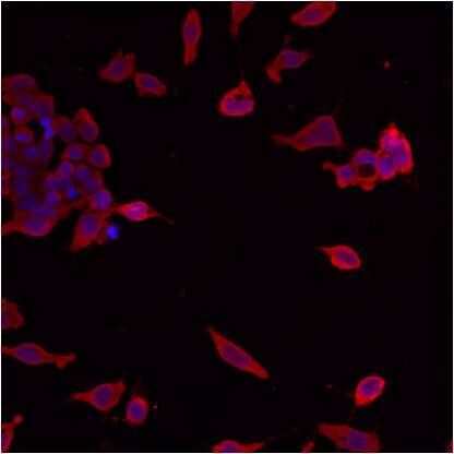
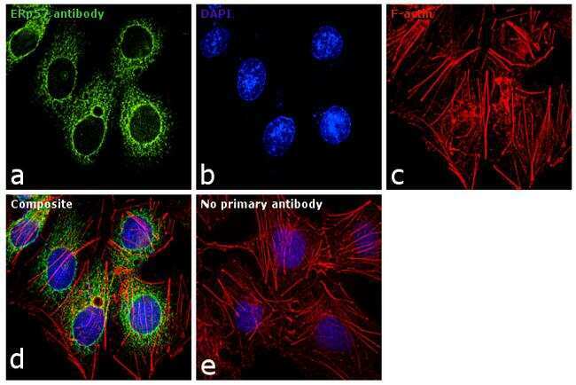
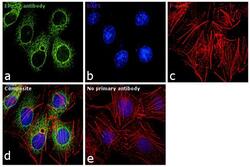
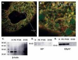
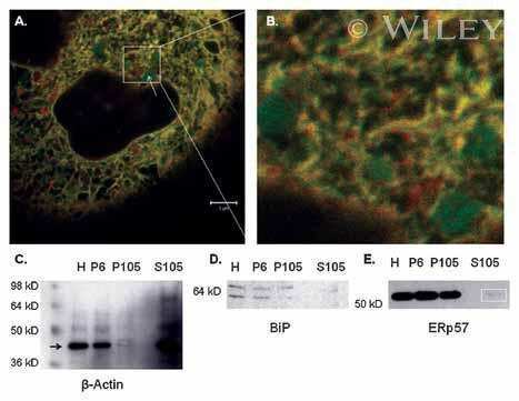
- Immunofluorescence analysis of ERp57 was performed using 70% confluent log phase HepG2 cells. The cells were fixed with 4% paraformaldehyde for 10 minutes, permeabilized with 0.1% Triton™ X-100 for 10 minutes, and blocked with 1% BSA for 1 hour at room temperature. The cells were labeled with ERp57 Rabbit Polyclonal Antibody (Product # PA3-009) at 1:250 in 0.1% BSA and incubated for overnight at 4 degree Celsius and then labeled with Goat anti-Rabbit IgG (H+L) Superclonal™ Secondary Antibody, Alexa Fluor® 488 conjugate (Product # A27034) at a dilution of 1:2000 for 45 minutes at room temperature (Panel a: green). Nuclei (Panel b: blue) were stained with SlowFade® Gold Antifade Mountant with DAPI (Product # S36938). F-actin (Panel c: red) was stained with Rhodamine Phalloidin (Product # R415, 1:300). Panel d represents the merged image showing cytoplasmic localization. Panel e shows the no primary antibody control. The images were captured at 60X magnification.
- Submitted by
- Invitrogen Antibodies (provider)
- Main image
- Experimental details
- Immunofluorescent analysis of ERp57 using anti-ERp57 polyclonal antibody (Product # PA3-009) shows staining in HMVEC Cells.

- Submitted by
- Invitrogen Antibodies (provider)
- Main image
- Experimental details
- Immunofluorescent analysis of ERp57 using anti-ERp57 polyclonal antibody (Product # PA3-009) shows staining in p19 Cells.

- Submitted by
- Invitrogen Antibodies (provider)
- Main image

- Experimental details
- Immunofluorescent analysis of ERp57 using anti-ERp57 polyclonal antibody (Product # PA3-009) shows staining in NS-1 Cells.
- Submitted by
- Invitrogen Antibodies (provider)
- Main image

- Experimental details
- Immunofluorescent analysis of ERp57 using anti-ERp57 polyclonal antibody (Product # PA3-009) shows staining in HMVEC Cells.
- Submitted by
- Invitrogen Antibodies (provider)
- Main image
- Experimental details
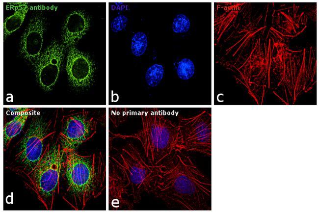
- Immunofluorescence analysis of ERp57 was performed using 70% confluent log phase HepG2 cells. The cells were fixed with 4% paraformaldehyde for 10 minutes, permeabilized with 0.1% Triton™ X-100 for 10 minutes, and blocked with 1% BSA for 1 hour at room temperature. The cells were labeled with ERp57 Rabbit Polyclonal Antibody (Product # PA3-009) at 1:250 in 0.1% BSA and incubated for overnight at 4 degree Celsius and then labeled with Goat anti-Rabbit IgG (Heavy Chain) Superclonal™ Secondary Antibody, Alexa Fluor® 488 conjugate (Product # A27034) at a dilution of 1:2000 for 45 minutes at room temperature (Panel a: green). Nuclei (Panel b: blue) were stained with SlowFade® Gold Antifade Mountant with DAPI (Product # S36938). F-actin (Panel c: red) was stained with Rhodamine Phalloidin (Product # R415, 1:300). Panel d represents the merged image showing cytoplasmic localization. Panel e shows the no primary antibody control. The images were captured at 60X magnification.
Supportive validation
- Submitted by
- Invitrogen Antibodies (provider)
- Main image

- Experimental details
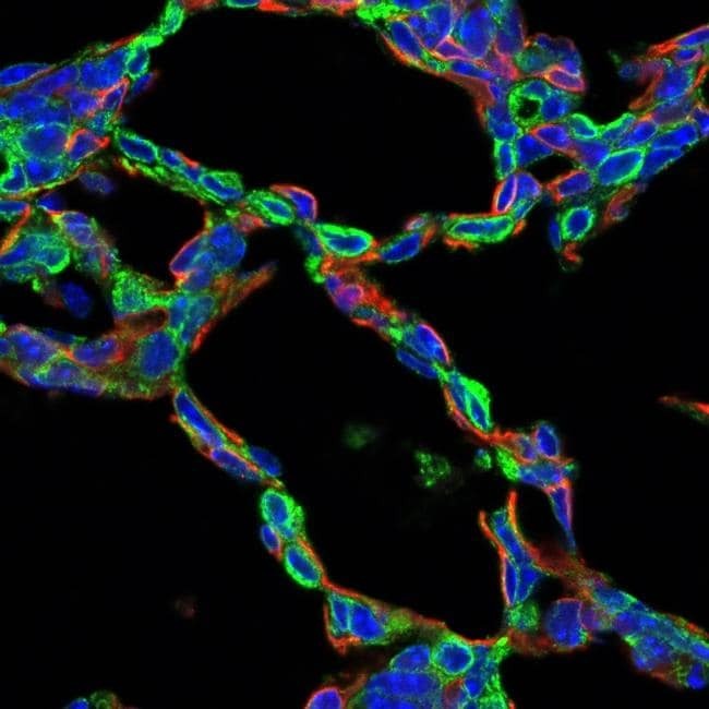
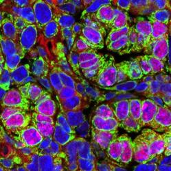
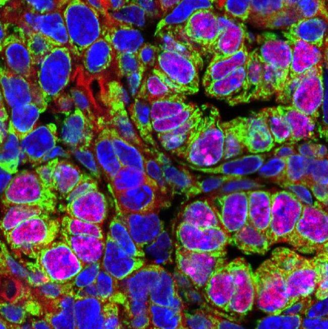
- Immunohistochemistry analysis of PDIA3 (ERp57) was performed on frozen sections of postnatal day 1 mouse lung tissue. To expose target proteins, antigen retrieval was performed using Citrate buffer, pH=6 using microwave. Following antigen retrieval, tissues were blocked in 4% Normal Donkey Serum (NDS) for 1-2 hours at room temperature. Tissue samples were then probed with anti-Pdia3/Erp57 antibody (Product # PA3-009) at a dilution of 1:200 (Green) along with anti-Emcn as endothelial cell marker (Red). Primary antibody incubation was carried out overnight at 4°C in a humidified chamber. Tissues were washed extensively with PBS/0.2% Triton X to remove unbound antibody. Detection was performed using Alexa Fluor conjugated anti-rabbit and anti-mouse IgG secondary antibodies at a dilution of 1:500. Following secondary antibody incubation tissues were washed extensively with PBS/0.2% Triton X and mounted using Prolong Gold anti-fade reagent (Product # P36930). Confocal images were taken on a Nikon A1 LUNA inverted microscope using 60X water or 100X oil objectives. Pdia3 is not expressed in endothelial cells based on non-colocalizaition with endomucin (emcn). Data courtesy of Dr. Jeff Whitsett's lab.
- Submitted by
- Invitrogen Antibodies (provider)
- Main image
- Experimental details
- Immunohistochemistry analysis of PDIA3 (ERp57) was performed on frozen sections of e18.5 mouse lung tissue. To expose target proteins, antigen retrieval was performed using Citrate buffer, pH=6 using microwave. Following antigen retrieval, tissues were blocked in 4% Normal Donkey Serum (NDS) for 1-2 hours at room temperature. Tissue samples were then probed with anti-Pdia3/Erp57 antibody (Product # PA3-009) at a dilution of 1:200 (Green) along with anti-Nkx2.1 as epithelial cell marker (Red). Primary antibody incubation was carried out overnight at 4°C in a humidified chamber. Tissues were washed extensively with PBS/0.2% Triton X to remove unbound antibody. Detection was performed using Alexa Fluor conjugated anti-rabbit and anti-mouse IgG secondary antibodies at a dilution of 1:500. Following secondary antibody incubation tissues were washed extensively with PBS/0.2% Triton X and mounted using Prolong Gold anti-fade reagent (Product # P36930). Confocal images were taken on a Nikon A1 LUNA inverted microscope using 60X water or 100X oil objectives. Pdia3 is localized in epithelial cells on co-localization with Nkx2.1. Data courtesy of Dr. Jeff Whitsett's lab.
Supportive validation
- Submitted by
- Invitrogen Antibodies (provider)
- Main image
- Experimental details
- NULL
- Submitted by
- Invitrogen Antibodies (provider)
- Main image

- Experimental details
- NULL

- Submitted by
- Invitrogen Antibodies (provider)
- Main image
- Experimental details
- NULL
- Submitted by
- Invitrogen Antibodies (provider)
- Main image
- Experimental details
- NULL
- Submitted by
- Invitrogen Antibodies (provider)
- Main image

- Experimental details
- NULL
- Submitted by
- Invitrogen Antibodies (provider)
- Main image
- Experimental details
- NULL