Explore
 Validate
 Learn
LearnNBP1-84796
antibody from Novus Biologicals
Targeting: PDIA3
ERp57, ERp60, ERp61, GRP57, GRP58, HsT17083, P58, PI-PLC
 Western blot
 Immunocytochemistry
Antibody data
- Antibody Data
- Antigen structure
- References [4]
- Comments [0]
- Validations
- Western blot [4]
- Immunohistochemistry [10]
Submit
Validation data
Reference
Comment
Report error
- Product number
- NBP1-84796 - Provider product page

- Provider
- Novus Biologicals
- Proper citation
- Novus Cat#NBP1-84796, RRID:AB_11022828
- Product name
- Rabbit Polyclonal ERp57/PDIA3 Antibody
- Antibody type
- Polyclonal
- Description
- Immunogen affinity purified. Specificity of human ERp57/PDIA3 antibody verified on a Protein Array containing target protein plus 383 other non-specific proteins.
- Reactivity
- Human, Mouse, Rat
- Host
- Rabbit
- Isotype
- IgG
- Vial size
- 0.1 ml
- Storage
- Store at 4C short term. Aliquot and store at -20C long term. Avoid freeze-thaw cycles.
Submitted references The dehydrogenase region of the NADPH oxidase component Nox2 acts as a protein disulfide isomerase (PDI) resembling PDIA3 with a role in the binding of the activator protein p67 (phox.).
Proteomics based identification of cell migration related proteins in HBV expressing HepG2 cells.
Loss-of-function mutations in MICU1 cause a brain and muscle disorder linked to primary alterations in mitochondrial calcium signaling.
The involvement of SMILE/TMTC3 in endoplasmic reticulum stress response.
Bechor E, Dahan I, Fradin T, Berdichevsky Y, Zahavi A, Federman Gross A, Rafalowski M, Pick E
Frontiers in chemistry 2015;3:3
Frontiers in chemistry 2015;3:3
Proteomics based identification of cell migration related proteins in HBV expressing HepG2 cells.
Feng H, Li X, Chan V, Chen WN
PloS one 2014;9(4):e95621
PloS one 2014;9(4):e95621
Loss-of-function mutations in MICU1 cause a brain and muscle disorder linked to primary alterations in mitochondrial calcium signaling.
Logan CV, Szabadkai G, Sharpe JA, Parry DA, Torelli S, Childs AM, Kriek M, Phadke R, Johnson CA, Roberts NY, Bonthron DT, Pysden KA, Whyte T, Munteanu I, Foley AR, Wheway G, Szymanska K, Natarajan S, Abdelhamed ZA, Morgan JE, Roper H, Santen GW, Niks EH, van der Pol WL, Lindhout D, Raffaello A, De Stefani D, den Dunnen JT, Sun Y, Ginjaar I, Sewry CA, Hurles M, Rizzuto R, UK10K Consortium., Duchen MR, Muntoni F, Sheridan E
Nature genetics 2014 Feb;46(2):188-93
Nature genetics 2014 Feb;46(2):188-93
The involvement of SMILE/TMTC3 in endoplasmic reticulum stress response.
Racapé M, Duong Van Huyen JP, Danger R, Giral M, Bleicher F, Foucher Y, Pallier A, Pilet P, Tafelmeyer P, Ashton-Chess J, Dugast E, Pettré S, Charreau B, Soulillou JP, Brouard S
PloS one 2011;6(5):e19321
PloS one 2011;6(5):e19321
No comments: Submit comment
Supportive validation
- Submitted by
- Novus Biologicals (provider)
- Main image
- Experimental details
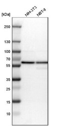
- Western Blot: ERp57/PDIA3 Antibody [NBP1-84796] - Analysis in mouse cell line NIH-3T3 and rat cell line NBT-II.

- Submitted by
- Novus Biologicals (provider)
- Main image

- Experimental details
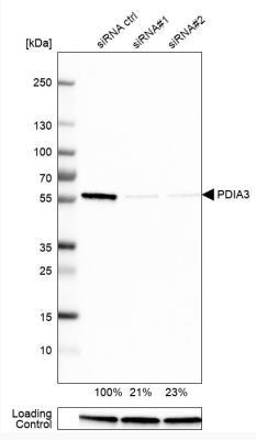
- Western Blot: ERp57/PDIA3 Antibody [NBP1-84796] - Analysis in U-251MG cells transfected with control siRNA, target specific siRNA probe #1 and #2, using Anti-PDIA3 antibody. Remaining relative intensity is presented. Loading control: Anti-GAPDH.
- Submitted by
- Novus Biologicals (provider)
- Main image
- Experimental details
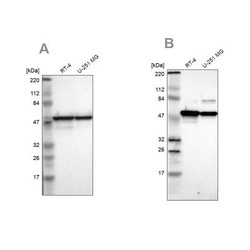
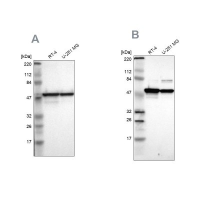
- Western Blot: ERp57/PDIA3 Antibody [NBP1-84796] - Analysis using Anti-PDIA3 antibody NBP1-84796 (A) shows similar pattern to independent antibody NBP1-84797 (B).
- Submitted by
- Novus Biologicals (provider)
- Main image
- Experimental details
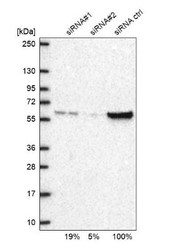
- Western Blot: ERp57/PDIA3 Antibody [NBP1-84796] - Analysis in U2OS cells transfected with control siRNA, target specific siRNA probe #1 and #2,. Remaining relative intensity is presented

Supportive validation
- Submitted by
- Novus Biologicals (provider)
- Main image

- Experimental details
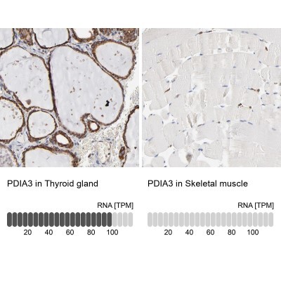
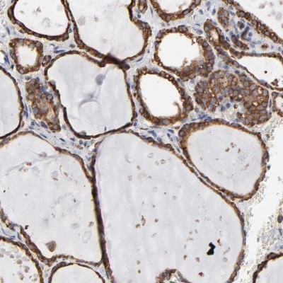
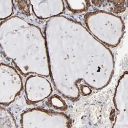
- Immunohistochemistry-Paraffin: ERp57/PDIA3 Antibody [NBP1-84796] - Staining of human thyroid gland shows strong cytoplasmic positivity in glandular cells.
- Submitted by
- Novus Biologicals (provider)
- Main image

- Experimental details
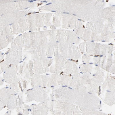
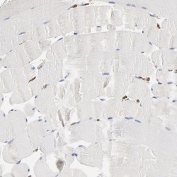
- Immunohistochemistry-Paraffin: ERp57/PDIA3 Antibody [NBP1-84796] - Staining of human skeletal muscle shows low expression as expected.
- Submitted by
- Novus Biologicals (provider)
- Main image
- Experimental details
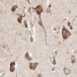
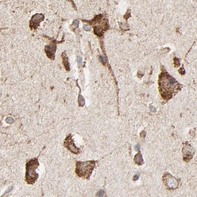
- Immunohistochemistry-Paraffin: ERp57/PDIA3 Antibody [NBP1-84796] - Staining of human cerebral cortex.
- Submitted by
- Novus Biologicals (provider)
- Main image
- Experimental details
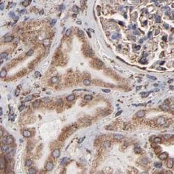
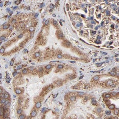
- Immunohistochemistry-Paraffin: ERp57/PDIA3 Antibody [NBP1-84796] - Staining of human kidney.
- Submitted by
- Novus Biologicals (provider)
- Main image

- Experimental details
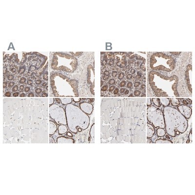
- Immunohistochemistry-Paraffin: ERp57/PDIA3 Antibody [NBP1-84796] - Staining of human gastrointestinal, prostate, skeletal muscle and thyroid gland using Anti-PDIA3 antibody NBP1-84796 (A) shows similar protein distribution across tissues to independent antibody NBP1-84797 (B).
- Submitted by
- Novus Biologicals (provider)
- Main image

- Experimental details
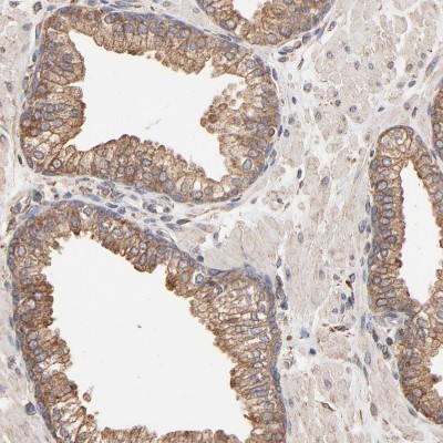
- Immunohistochemistry-Paraffin: ERp57/PDIA3 Antibody [NBP1-84796] - Staining of human prostate shows moderate cytoplasmic positivity in glandular cells.
- Submitted by
- Novus Biologicals (provider)
- Main image
- Experimental details
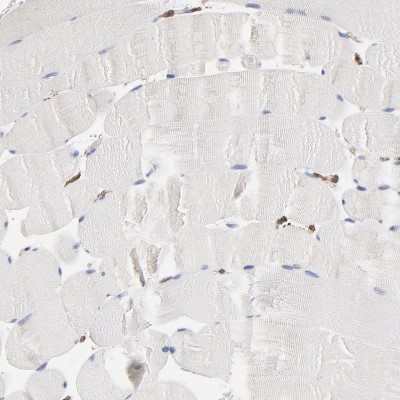
- Immunohistochemistry-Paraffin: ERp57/PDIA3 Antibody [NBP1-84796] - Staining of human skeletal muscle shows no cytoplasmic positivity in myocytes as expected.
- Submitted by
- Novus Biologicals (provider)
- Main image
- Experimental details
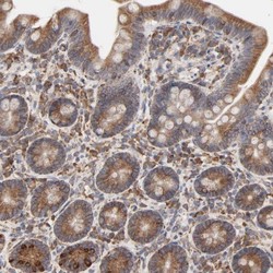
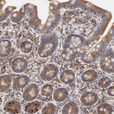
- Immunohistochemistry-Paraffin: ERp57/PDIA3 Antibody [NBP1-84796] - Staining of human small intestine shows moderate to strong cytoplasmic positivity in glandular cells.
- Submitted by
- Novus Biologicals (provider)
- Main image
- Experimental details
- Immunohistochemistry-Paraffin: ERp57/PDIA3 Antibody [NBP1-84796] - Staining of human thyroid gland shows strong cytoplasmic positivity in glandular cells.

- Submitted by
- Novus Biologicals (provider)
- Main image
- Experimental details
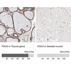
- Immunohistochemistry-Paraffin: ERp57/PDIA3 Antibody [NBP1-84796] - Staining in human thyroid gland and skeletal muscle tissues using NBP1-84796 antibody. Corresponding PDIA3 RNA-seq data are presented for the same tissues.