Explore
 Validate
 Learn
LearnHPA002645
antibody from Atlas Antibodies
Targeting: PDIA3
ERp57, ERp60, ERp61, GRP57, GRP58, HsT17083, P58, PI-PLC
 Western blot
 Immunocytochemistry
Antibody data
- Antibody Data
- Antigen structure
- References [2]
- Comments [0]
- Validations
- Western blot [1]
- Immunocytochemistry [1]
- Immunohistochemistry [1]
Submit
Validation data
Reference
Comment
Report error
- Product number
- HPA002645 - Provider product page

- Provider
- Atlas Antibodies
- Proper citation
- Atlas Antibodies Cat#HPA002645, RRID:AB_1079031
- Product name
- Anti-PDIA3
- Antibody type
- Polyclonal
- Description
- Polyclonal Antibody against Human PDIA3, Gene description: protein disulfide isomerase family A, member 3, Alternative Gene Names: ERp57, ERp60, ERp61, GRP57, GRP58, HsT17083, P58, PI-PLC, Validated applications: IHC, ICC, WB, Uniprot ID: P30101, Storage: Store at +4°C for short term storage. Long time storage is recommended at -20°C.
- Reactivity
- Human
- Host
- Rabbit
- Conjugate
- Unconjugated
- Isotype
- IgG
- Vial size
- 100 µl
- Concentration
- 0.1 mg/ml
- Storage
- Store at +4°C for short term storage. Long time storage is recommended at -20°C.
- Handling
- The antibody solution should be gently mixed before use.
Submitted references P4HB and PDIA3 are associated with tumor progression and therapeutic outcome of diffuse gliomas
SILAC-based quantitative proteomic approach to identify potential biomarkers from the esophageal squamous cell carcinoma secretome
Zou H, Wen C, Peng Z, Shao Y, Hu L, Li S, Li C, Zhou H
Oncology Reports 2017
Oncology Reports 2017
SILAC-based quantitative proteomic approach to identify potential biomarkers from the esophageal squamous cell carcinoma secretome
Kashyap M, Harsha H, Renuse S, Pawar H, Sahasrabuddhe N, Kim M, Marimuthu A, Keerthikumar S, Muthusamy B, Kandasamy K, Subbannayya Y, Prasad T, Mahmood R, Chaerkady R, Meltzer S, Kumar R, Rustgi A, Pandey A
Cancer Biology & Therapy 2014;10(8):796-810
Cancer Biology & Therapy 2014;10(8):796-810
No comments: Submit comment
Enhanced validation
- Submitted by
- Atlas Antibodies (provider)
- Enhanced method
- Genetic validation
- Main image
- Experimental details
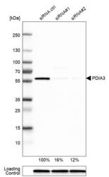
- Western blot analysis in U-251MG cells transfected with control siRNA, target specific siRNA probe #1 and #2, using Anti-PDIA3 antibody. Remaining relative intensity is presented. Loading control: Anti-GAPDH.
- Sample type
- Human
- Protocol
- Protocol

Supportive validation
- Submitted by
- Atlas Antibodies (provider)
- Main image

- Experimental details
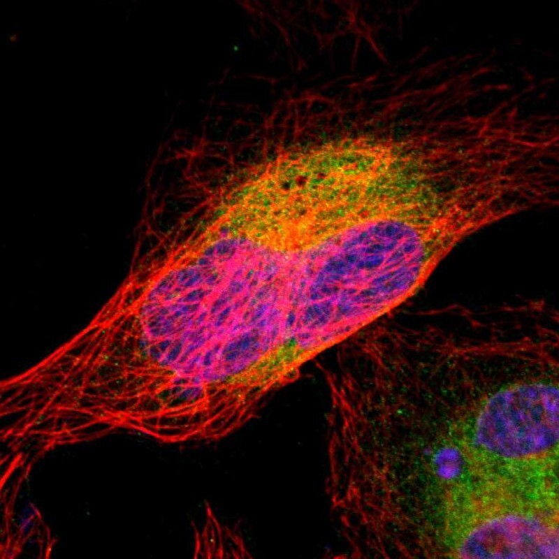
- Immunofluorescent staining of human cell line U-2 OS shows localization to endoplasmic reticulum.
- Sample type
- Human
Supportive validation
- Submitted by
- Atlas Antibodies (provider)
- Enhanced method
- Orthogonal validation
- Main image

- Experimental details
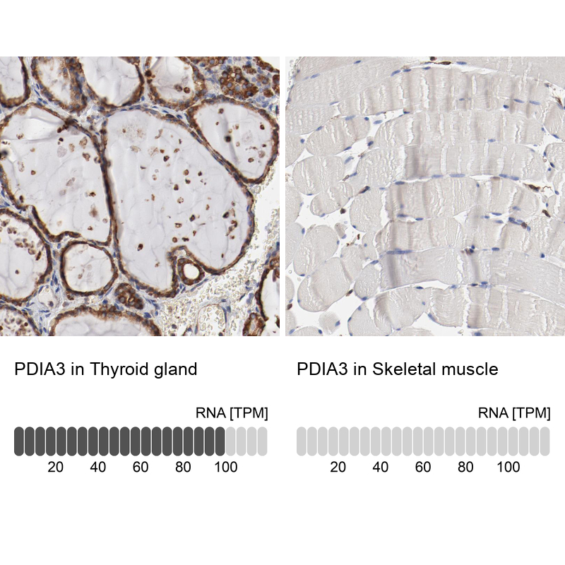
- Immunohistochemistry analysis in human thyroid gland and skeletal muscle tissues using HPA002645 antibody. Corresponding PDIA3 RNA-seq data are presented for the same tissues.
- Sample type
- Human
- Protocol
- Protocol