Explore
 Validate
 Learn
LearnHPA003230
antibody from Atlas Antibodies
Targeting: PDIA3
ERp57, ERp60, ERp61, GRP57, GRP58, HsT17083, P58, PI-PLC
 Western blot
 Immunohistochemistry
Antibody data
- Antibody Data
- Antigen structure
- References [8]
- Comments [0]
- Validations
- Western blot [1]
- Immunocytochemistry [2]
- Immunohistochemistry [1]
Submit
Validation data
Reference
Comment
Report error
- Product number
- HPA003230 - Provider product page

- Provider
- Atlas Antibodies
- Proper citation
- Atlas Antibodies Cat#HPA003230, RRID:AB_1079030
- Product name
- Anti-PDIA3
- Antibody type
- Polyclonal
- Description
- Polyclonal Antibody against Human PDIA3, Gene description: protein disulfide isomerase family A, member 3, Alternative Gene Names: ERp57, ERp60, ERp61, GRP57, GRP58, HsT17083, P58, PI-PLC, Validated applications: IHC, ICC, WB, Uniprot ID: P30101, Storage: Store at +4°C for short term storage. Long time storage is recommended at -20°C.
- Reactivity
- Human, Mouse, Rat
- Host
- Rabbit
- Conjugate
- Unconjugated
- Isotype
- IgG
- Vial size
- 100 µl
- Concentration
- 0.3 mg/ml
- Storage
- Store at +4°C for short term storage. Long time storage is recommended at -20°C.
- Handling
- The antibody solution should be gently mixed before use.
Submitted references Robust CXCL10/IP-10 and CCL5/RANTES Production Induced by Tick-Borne Encephalitis Virus in Human Brain Pericytes Despite Weak Infection
Loss of surface transport is a main cellular pathomechanism of CRB2 variants causing podocytopathies
Inhibition of calpain 1 restores plasma membrane stability to pharmacologically rescued Phe508del-CFTR variant
Characterization of Ηeparan Sulfate Proteoglycan-positive Recycling Endosomes Isolated from Glioma Cells
The dehydrogenase region of the NADPH oxidase component Nox2 acts as a protein disulfide isomerase (PDI) resembling PDIA3 with a role in the binding of the activator protein p67phox
Proteomics Based Identification of Cell Migration Related Proteins in HBV Expressing HepG2 Cells
Loss-of-function mutations in MICU1 cause a brain and muscle disorder linked to primary alterations in mitochondrial calcium signaling
The Involvement of SMILE/TMTC3 in Endoplasmic Reticulum Stress Response
Prančlová V, Hönig V, Zemanová M, Růžek D, Palus M
International Journal of Molecular Sciences 2024;25(14):7892
International Journal of Molecular Sciences 2024;25(14):7892
Loss of surface transport is a main cellular pathomechanism of CRB2 variants causing podocytopathies
Möller-Kerutt A, Schönhoff B, Rellmann Y, George B, Braun D, Pavenstädt H, Weide T
Life Science Alliance 2023;6(3):e202201649
Life Science Alliance 2023;6(3):e202201649
Inhibition of calpain 1 restores plasma membrane stability to pharmacologically rescued Phe508del-CFTR variant
Matos A, Pinto F, Barros P, Amaral M, Pepperkok R, Matos P
Journal of Biological Chemistry 2019;294(36):13396-13410
Journal of Biological Chemistry 2019;294(36):13396-13410
Characterization of Ηeparan Sulfate Proteoglycan-positive Recycling Endosomes Isolated from Glioma Cells
PODYMA-INOUE, K, MORIWAKI T, RAJAPAKSHE A, TERASAWA K, HARA-YOKOYAMA M
Cancer Genomics & Proteomics 2016;13(6):443-452
Cancer Genomics & Proteomics 2016;13(6):443-452
The dehydrogenase region of the NADPH oxidase component Nox2 acts as a protein disulfide isomerase (PDI) resembling PDIA3 with a role in the binding of the activator protein p67phox
Bechor E, Dahan I, Fradin T, Berdichevsky Y, Zahavi A, Federman Gross A, Rafalowski M, Pick E
Frontiers in Chemistry 2015;3
Frontiers in Chemistry 2015;3
Proteomics Based Identification of Cell Migration Related Proteins in HBV Expressing HepG2 Cells
Kaushik-Basu N, Feng H, Li X, Chan V, Chen W
PLoS ONE 2014;9(4):e95621
PLoS ONE 2014;9(4):e95621
Loss-of-function mutations in MICU1 cause a brain and muscle disorder linked to primary alterations in mitochondrial calcium signaling
Logan C, Szabadkai G, Sharpe J, Parry D, Torelli S, Childs A, Kriek M, Phadke R, Johnson C, Roberts N, Bonthron D, Pysden K, Whyte T, Munteanu I, Foley A, Wheway G, Szymanska K, Natarajan S, Abdelhamed Z, Morgan J, Roper H, Santen G, Niks E, van der Pol W, Lindhout D, Raffaello A, De Stefani D, den Dunnen J, Sun Y, Ginjaar I, Sewry C, Hurles M, Rizzuto R, Duchen M, Muntoni F, Sheridan E
Nature Genetics 2013;46(2):188-193
Nature Genetics 2013;46(2):188-193
The Involvement of SMILE/TMTC3 in Endoplasmic Reticulum Stress Response
Câmara N, Racapé M, Duong Van Huyen J, Danger R, Giral M, Bleicher F, Foucher Y, Pallier A, Pilet P, Tafelmeyer P, Ashton-Chess J, Dugast E, Pettré S, Charreau B, Soulillou J, Brouard S
PLoS ONE 2011;6(5):e19321
PLoS ONE 2011;6(5):e19321
No comments: Submit comment
Enhanced validation
- Submitted by
- Atlas Antibodies (provider)
- Enhanced method
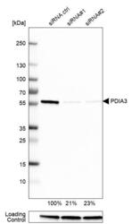
- Genetic validation
- Main image
- Experimental details
- Western blot analysis in U-251MG cells transfected with control siRNA, target specific siRNA probe #1 and #2, using Anti-PDIA3 antibody. Remaining relative intensity is presented. Loading control: Anti-GAPDH.
- Sample type
- Human
- Protocol
- Protocol

Enhanced validation
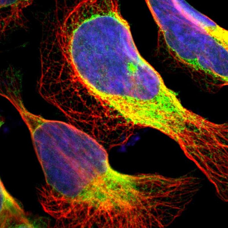
Supportive validation
- Submitted by
- 55af80e3e0991
- Enhanced method
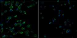
- Genetic validation
- Main image
- Experimental details
- Confocal images of immunofluorescently stained human U-2 OS cells.The protein PDIA3 is shown in green and the nucleus in blue. The image to the left show cells transfected with control siRNA and the image to the right show cells where PDIA3 has been downregulated with specific siRNA.
- Sample type
- U-2 OS cells
- Primary Ab dilution
- 1:127
- Secondary Ab
- Secondary Ab
- Secondary Ab dilution
- 1:800
- Knockdown/Genetic Approaches Application
- Immunocytochemistry
Supportive validation
- Submitted by
- Atlas Antibodies (provider)
- Main image
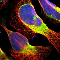
- Experimental details
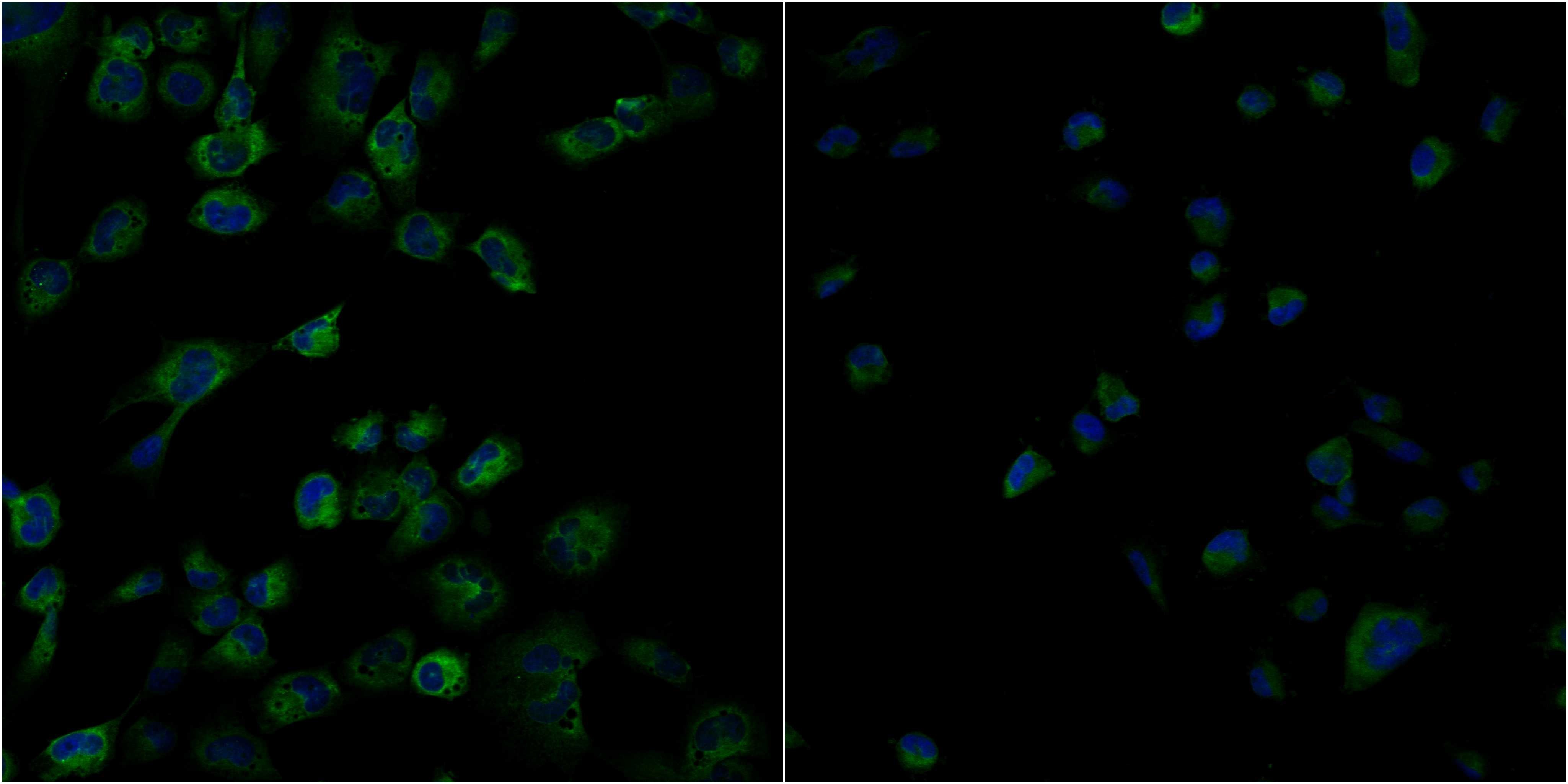
- Immunofluorescent staining of human cell line U-2 OS shows localization to endoplasmic reticulum.
- Sample type
- Human
Supportive validation
- Submitted by
- Atlas Antibodies (provider)
- Enhanced method
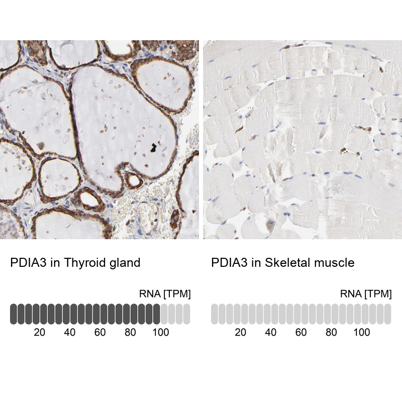
- Orthogonal validation
- Main image

- Experimental details
- Immunohistochemistry analysis in human thyroid gland and skeletal muscle tissues using HPA003230 antibody. Corresponding PDIA3 RNA-seq data are presented for the same tissues.
- Sample type
- Human
- Protocol
- Protocol