Explore
 Validate
 Learn
Learn Western blot
Antibody data
- Antibody Data
- Antigen structure
- References [16]
- Comments [0]
- Validations
- Western blot [2]
- Immunocytochemistry [3]
- Immunohistochemistry [4]
Submit
Validation data
Reference
Comment
Report error
- Product number
- MA5-13144 - Provider product page

- Provider
- Invitrogen Antibodies
- Product name
- Cytokeratin Pan Type I Monoclonal Antibody (AE1)
- Antibody type
- Monoclonal
- Antigen
- Other
- Description
- MA5-13144 targets Cytokeratin Low Molecular Weight in WB, IF and IHC (P) applications and shows reactivity with Bovine, Chicken, Human, mouse, Non-human primate, Rabbit, and Rat samples. This antibody is not recommended for NIH-3T3 cells in immunofluorescent applications.
- Antibody clone number
- AE1
- Concentration
- 0.2 mg/mL
Submitted references Fumarate induces redox-dependent senescence by modifying glutathione metabolism.
Association of Merkel cell polyomavirus infection with EGFR mutation status in Chinese non-small cell lung cancer patients.
A clinicopathological study of the significance of the proportion of choroid morphology in chordoid meningioma.
The prognostic impact of O6-methylguanine DNA methyltransferase and epidermal growth factor receptor expressions on primary gliosarcoma: a clinicopathologic and immunohistochemical study of seven cases at a single institution.
Clear cell meningioma with frequent chordoid features and aggressive behavior: a clinicopathologic study of ten cases at a single institution.
Novel immunofluorescence protocol for multimarker assessment of putative disseminating breast cancer stem cells.
Rhabdoid papillary meningioma: a clinicopathologic case series study.
Chordoid meningioma: a clinicopathologic study of 11 cases at a single institution.
Assessment of the tumorigenesis and drug susceptibility of three new canine mammary tumor cell lines.
An immunohistochemical study of feline endometrial adenocarcinoma.
Coexistence of different tissue tumourigenesis in an N-methyl-N-nitrosourea-induced mammary carcinoma model: a histopathological report in Sprague-Dawley rats.
Melanotic Xp11 translocation renal cancer: a case with PSF-TFE3 gene fusion and up-regulation of melanogenetic transcripts.
Detection of micrometastases and skip metastases with ex vivo sentinel node mapping in carcinoma of the colon and rectum.
Mediastinal intraoperative radioisotope sentinel lymph node mapping in non-small-cell lung cancer.
Possible association between HPV16 E7 protein level and cytokeratin 19.
Ectopic thyroid in the gallbladder: report of a case.
Zheng L, Cardaci S, Jerby L, MacKenzie ED, Sciacovelli M, Johnson TI, Gaude E, King A, Leach JD, Edrada-Ebel R, Hedley A, Morrice NA, Kalna G, Blyth K, Ruppin E, Frezza C, Gottlieb E
Nature communications 2015 Jan 23;6:6001
Nature communications 2015 Jan 23;6:6001
Association of Merkel cell polyomavirus infection with EGFR mutation status in Chinese non-small cell lung cancer patients.
Xu S, Jiang J, Yu X, Sheng D, Zhu T, Jin M
Lung cancer (Amsterdam, Netherlands) 2014 Mar;83(3):341-6
Lung cancer (Amsterdam, Netherlands) 2014 Mar;83(3):341-6
A clinicopathological study of the significance of the proportion of choroid morphology in chordoid meningioma.
Lin JW, Lu CH, Lin WC, Wu YT, Huang YJ, Shih FY, Ho JT, Chuang MJ
Journal of clinical neuroscience : official journal of the Neurosurgical Society of Australasia 2012 Jun;19(6):836-43
Journal of clinical neuroscience : official journal of the Neurosurgical Society of Australasia 2012 Jun;19(6):836-43
The prognostic impact of O6-methylguanine DNA methyltransferase and epidermal growth factor receptor expressions on primary gliosarcoma: a clinicopathologic and immunohistochemical study of seven cases at a single institution.
Lin JW, Wu YT, Chang IW
Indian journal of pathology & microbiology 2011 Oct-Dec;54(4):683-7
Indian journal of pathology & microbiology 2011 Oct-Dec;54(4):683-7
Clear cell meningioma with frequent chordoid features and aggressive behavior: a clinicopathologic study of ten cases at a single institution.
Chen HK, Wu YT, Lin YJ, Lin JW
Journal of neuro-oncology 2011 Jul;103(3):551-9
Journal of neuro-oncology 2011 Jul;103(3):551-9
Novel immunofluorescence protocol for multimarker assessment of putative disseminating breast cancer stem cells.
Balic M, Rapp N, Stanzer S, Lin H, Strutz J, Szkandera J, Daidone MG, Samonigg H, Cote RJ, Dandachi N
Applied immunohistochemistry & molecular morphology : AIMM 2011 Jan;19(1):33-40
Applied immunohistochemistry & molecular morphology : AIMM 2011 Jan;19(1):33-40
Rhabdoid papillary meningioma: a clinicopathologic case series study.
Wu YT, Ho JT, Lin YJ, Lin JW
Neuropathology : official journal of the Japanese Society of Neuropathology 2011 Dec;31(6):599-605
Neuropathology : official journal of the Japanese Society of Neuropathology 2011 Dec;31(6):599-605
Chordoid meningioma: a clinicopathologic study of 11 cases at a single institution.
Lin JW, Ho JT, Lin YJ, Wu YT
Journal of neuro-oncology 2010 Dec;100(3):465-73
Journal of neuro-oncology 2010 Dec;100(3):465-73
Assessment of the tumorigenesis and drug susceptibility of three new canine mammary tumor cell lines.
Chang CY, Chiou PP, Chen WJ, Li YH, Yiu JC, Cheng YH, Chen SD, Lin CT, Lai YS
Research in veterinary science 2010 Apr;88(2):285-93
Research in veterinary science 2010 Apr;88(2):285-93
An immunohistochemical study of feline endometrial adenocarcinoma.
Gil da Costa RM, Santos M, Amorim I, Lopes C, Pereira PD, Faustino AM
Journal of comparative pathology 2009 May;140(4):254-9
Journal of comparative pathology 2009 May;140(4):254-9
Coexistence of different tissue tumourigenesis in an N-methyl-N-nitrosourea-induced mammary carcinoma model: a histopathological report in Sprague-Dawley rats.
Esendagli G, Yilmaz G, Canpinar H, Gunel-Ozcan A, Guc MO, Guc D
Laboratory animals 2009 Jan;43(1):60-4
Laboratory animals 2009 Jan;43(1):60-4
Melanotic Xp11 translocation renal cancer: a case with PSF-TFE3 gene fusion and up-regulation of melanogenetic transcripts.
Chang IW, Huang HY, Sung MT
The American journal of surgical pathology 2009 Dec;33(12):1894-901
The American journal of surgical pathology 2009 Dec;33(12):1894-901
Detection of micrometastases and skip metastases with ex vivo sentinel node mapping in carcinoma of the colon and rectum.
Yagci G, Unlu A, Kurt B, Can MF, Kaymakcioglu N, Cetiner S, Tufan T, Sen D
International journal of colorectal disease 2007 Feb;22(2):167-73
International journal of colorectal disease 2007 Feb;22(2):167-73
Mediastinal intraoperative radioisotope sentinel lymph node mapping in non-small-cell lung cancer.
Atinkaya C, Ozlem Küçük N, Koparal H, Aras G, Sak SD, Ozdemir N
Nuclear medicine communications 2005 Aug;26(8):717-20
Nuclear medicine communications 2005 Aug;26(8):717-20
Possible association between HPV16 E7 protein level and cytokeratin 19.
Favia G, Kanduc D, Lo Muzio L, Lucchese A, Serpico R
International journal of cancer 2004 Sep 20;111(5):795-7
International journal of cancer 2004 Sep 20;111(5):795-7
Ectopic thyroid in the gallbladder: report of a case.
Ihtiyar E, Isiksoy S, Algin C, Sahin A, Erkasap S, Yasar B
Surgery today 2003;33(10):777-80
Surgery today 2003;33(10):777-80
No comments: Submit comment
Supportive validation
- Submitted by
- Invitrogen Antibodies (provider)
- Main image

- Experimental details
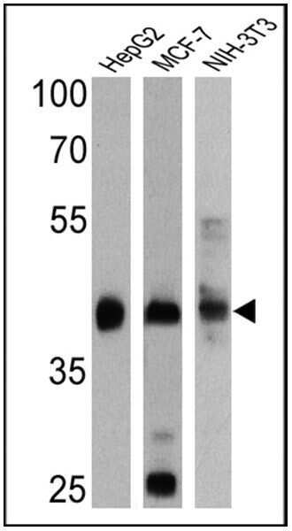
- Western blot analysis of Cytokeratin Low Molecular Weight was performed by loading 25 µg of HepG2 (Lane 1), MCF-7 (Lane 2) and NIH-3T3 (Lane 3) cell lysates onto an SDS polyacrylamide gel. Proteins were transferred to a PVDF membrane and blocked at 4ºC overnight. The membrane was probed with a Cytokeratin Low Molecular Weight monoclonal antibody (Product # MA5-13144) at a dilution of 1:4000 (HepG2 and MCF-7) and 1:1000 (NIH-3T3) overnight at 4°C, washed in TBST, and probed with an HRP-conjugated secondary antibody for 1 hr at room temperature in the dark. Chemiluminescent detection was performed using Pierce ECL Plus Western Blotting Substrate (Product # 32132). Results show a band at approx. 40 kDa.
- Submitted by
- Invitrogen Antibodies (provider)
- Main image

- Experimental details
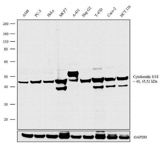
- Western blot analysis was performed on whole cell extracts (30 µg lysate) of A549 (Lane 1), PC-3 (Lane 2), HeLa (Lane 3), MCF7 (Lane 4), A-431 (Lane 5), Hep G2 (Lane 6), T-47D (Lane 7), Caco-2 (Lane 8) and HCT 116 (Lane 9). The blots were probed with Anti-Cytokeratin 8/18 (Cytokeratin Low Molecular Weight) Monoclonal Antibody (Product # MA5-13144, 1:2000 dilution) and detected by chemiluminescence using Goat anti-Mouse IgG (H+L) Superclonal™ Secondary Antibody, HRP conjugate (Product # A28177, 0.25 µg/mL, 1:4000 dilution). A 40 kDa, 45 kDa and 52 kDa band corresponding to Cytokeratin low molecular weight were observed across the cell lines tested. Known quantity of protein samples were electrophoresed using Novex® NuPAGE® 4-12 % Bis-Tris gel (Product # NP0321BOX), XCell SureLock™ Electrophoresis System (Product # EI0002) and Novex® Sharp Pre-Stained Protein Standard (Product # LC5800). Resolved proteins were then transferred onto a nitrocellulose membrane with iBlot® 2 Dry Blotting System (Product # IB21001). The membrane was probed with the relevant primary antibody following blocking with 5 % skimmed milk. Chemiluminescent detection was performed using Pierce™ ECL Western Blotting Substrate (Product # 32106).
Supportive validation
- Submitted by
- Invitrogen Antibodies (provider)
- Main image

- Experimental details
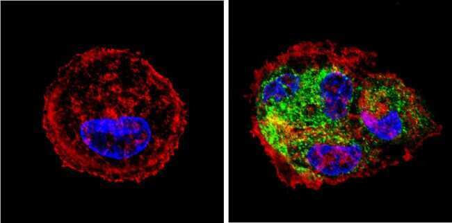
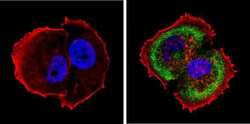
- Immunofluorescent analysis of Cytokeratin Low Molecular Weight (green) showing staining in the cytoplasm of HepG2 cells. Formalin-fixed cells were permeabilized with 0.1% Triton X-100 in TBS for 5-10 minutes and blocked with 3% BSA-PBS for 30 minutes at room temperature. Cells were probed with a Cytokeratin Low Molecular Weight monoclonal antibody (Product # MA5-13144) in 3% BSA-PBS at a dilution of 1:200 and incubated overnight at 4 ºC in a humidified chamber. Cells were washed with PBST and incubated with a DyLight-conjugated secondary antibody in PBS at room temperature in the dark. F-actin (red) was stained with a fluorescent red phalloidin and nuclei (blue) were stained with Hoechst or DAPI. Images were taken at a magnification of 60x.
- Submitted by
- Invitrogen Antibodies (provider)
- Main image
- Experimental details
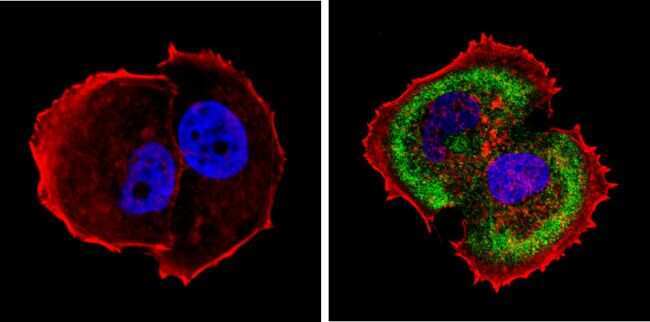
- Immunofluorescent analysis of Cytokeratin Low Molecular Weight (green) showing staining in the cytoplasm of MCF-7 cells. Formalin-fixed cells were permeabilized with 0.1% Triton X-100 in TBS for 5-10 minutes and blocked with 3% BSA-PBS for 30 minutes at room temperature. Cells were probed with a Cytokeratin Low Molecular Weight monoclonal antibody (Product # MA5-13144) in 3% BSA-PBS at a dilution of 1:200 and incubated overnight at 4 ºC in a humidified chamber. Cells were washed with PBST and incubated with a DyLight-conjugated secondary antibody in PBS at room temperature in the dark. F-actin (red) was stained with a fluorescent red phalloidin and nuclei (blue) were stained with Hoechst or DAPI. Images were taken at a magnification of 60x.
- Submitted by
- Invitrogen Antibodies (provider)
- Main image
- Experimental details
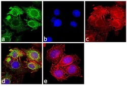
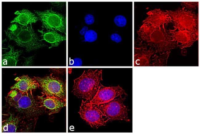
- Immunofluorescence analysis of Cytokeratin Low Molecular Weight was performed using 70% confluent log phase MCF7 cells. The cells were fixed with 4% paraformaldehyde for 10 minutes, permeabilized with 0.1% Triton™ X-100 for 10 minutes, and blocked with 1% BSA for 1 hour at room temperature. The cells were labeled with Cytokeratin 8/18 Monoclonal Antibody (Product # MA5-13144) at 5µg/mL in 0.1% BSA and incubated for 3 hours at room temperature and then labeled with Goat anti-Mouse IgG (H+L) Superclonal™ Secondary Antibody, Alexa Fluor® 488 conjugate (Product # A28175) at a dilution of 1:2000 for 45 minutes at room temperature (Panel a: green). Nuclei (Panel b: blue) were stained with SlowFade® Gold Antifade Mountant with DAPI (Product # S36938). F-actin (Panel c: red) was stained with Rhodamine Phalloidin (Product # R415, 1:300). Panel d represents the merged image showing cytoplasmic localization. Panel e shows the no primary antibody control. The images were captured at 60X magnification.
Supportive validation
- Submitted by
- Invitrogen Antibodies (provider)
- Main image

- Experimental details
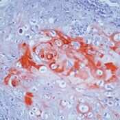
- Formalin-fixed, paraffin-embedded human squamous cell carcinoma of lung stained with Keratin, LMW antibody using peroxidase-conjugate and AEC chromogen. Note cytoplasmic staining of tumor cells.

- Submitted by
- Invitrogen Antibodies (provider)
- Main image

- Experimental details
- Formalin-fixed, paraffin-embedded human squamous cell carcinoma of lung stained with Keratin, LMW antibody using peroxidase-conjugate and AEC chromogen. Note cytoplasmic staining of tumor cells.
- Submitted by
- Invitrogen Antibodies (provider)
- Main image
- Experimental details
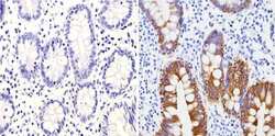
- Immunohistochemistry analysis of Cytokeratin Low Molecular Weight showing positive staining in the cytoplasm of paraffin-treated Human colon tissue (right) compared with a negative control in the absence of primary antibody (left). To expose target proteins, antigen retrieval method was performed using 10mM sodium citrate (pH 6.0), microwaved for 8-15 min. Following antigen retrieval, tissues were blocked in 3% H2O2-methanol for 15 min at room temperature, washed with ddH2O and PBS, and then probed with a Cytokeratin Low Molecular Weight monoclonal antibody (Product # MA5-13144) diluted by 3% BSA-PBS at a dilution of 1:200 overnight at 4°C in a humidified chamber. Tissues were washed extensively PBST and detection was performed using an HRP-conjugated secondary antibody followed by colorimetric detection using a DAB kit. Tissues were counterstained with hematoxylin and dehydrated with ethanol and xylene to prep for mounting.
- Submitted by
- Invitrogen Antibodies (provider)
- Main image

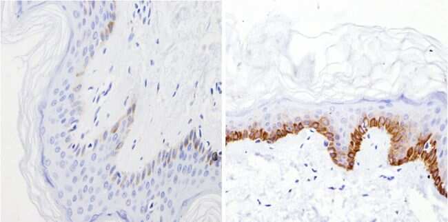
- Experimental details
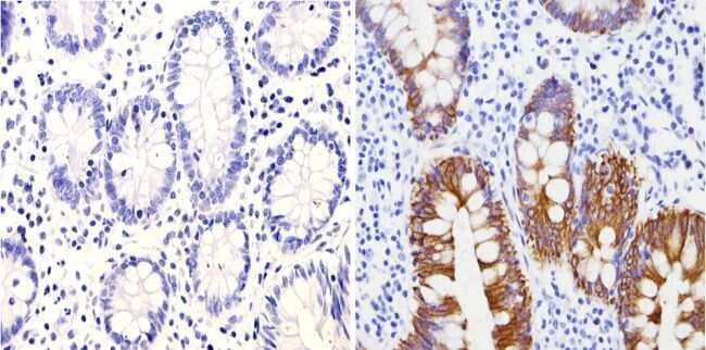
- Immunohistochemistry analysis of Cytokeratin Low Molecular Weight showing positive staining in the cytoplasm of paraffin-treated Human skin tissue (right) compared with a negative control in the absence of primary antibody (left). To expose target proteins, antigen retrieval method was performed using 10mM sodium citrate (pH 6.0), microwaved for 8-15 min. Following antigen retrieval, tissues were blocked in 3% H2O2-methanol for 15 min at room temperature, washed with ddH2O and PBS, and then probed with a Cytokeratin Low Molecular Weight monoclonal antibody (Product # MA5-13144) diluted by 3% BSA-PBS at a dilution of 1:200 overnight at 4°C in a humidified chamber. Tissues were washed extensively PBST and detection was performed using an HRP-conjugated secondary antibody followed by colorimetric detection using a DAB kit. Tissues were counterstained with hematoxylin and dehydrated with ethanol and xylene to prep for mounting.