Explore
 Validate
 Learn
Learn Western blot
Antibody data
- Antibody Data
- Antigen structure
- References [0]
- Comments [0]
- Validations
- Western blot [1]
- Immunocytochemistry [1]
- Immunohistochemistry [4]
- Other assay [1]
Submit
Validation data
Reference
Comment
Report error
- Product number
- MAB35061-100 - Provider product page

- Provider
- R&D Systems
- Product name
- Human Cytokeratin 19 Antibody
- Antibody type
- Monoclonal
- Description
- Protein A or G purified from hybridoma culture supernatant. Detects human Cytokeratin 19 in direct ELISAs and Western blots.
- Reactivity
- Human
- Host
- Mouse
- Conjugate
- Unconjugated
- Antigen sequence
P08727- Isotype
- IgG
- Antibody clone number
- 963420
- Vial size
- 100 ug
- Storage
- Use a manual defrost freezer and avoid repeated freeze-thaw cycles. 12 months from date of receipt, -20 to -70 °C as supplied. 1 month, 2 to 8 °C under sterile conditions after reconstitution. 6 months, -20 to -70 °C under sterile conditions after reconstitution.
No comments: Submit comment
Supportive validation
- Submitted by
- R&D Systems (provider)
- Main image
- Experimental details
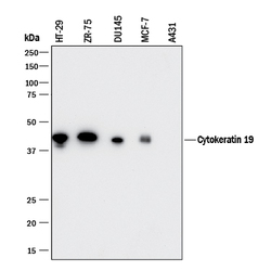
- Detection of Human Cytokeratin 19 by Western Blot. Western blot shows lysates of HT-29 human colon adenocarcinoma cell line, ZR-75 human breast cancer cell line, DU145 human prostate carcinoma cell line, MCF-7 human breast cancer cell line, and A431 human epithelial carcinoma cell line. PVDF membrane was probed with 0.25 µg/mL of Mouse Anti-Human Cytokeratin 19 Monoclonal Antibody (Catalog # MAB35061) followed by HRP-conjugated Anti-Mouse IgG Secondary Antibody (Catalog # HAF018). A specific band was detected for Cytokeratin 19 at approximately 40 kDa (as indicated). This experiment was conducted under reducing conditions and using Immunoblot Buffer Group 1.
Supportive validation
- Submitted by
- R&D Systems (provider)
- Main image

- Experimental details
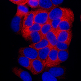
- Cytokeratin 19 in MCF-7 Human Cell Line. Cytokeratin 19 was detected in immersion fixed MCF-7 human breast cancer cell line using Mouse Anti-Human Cytokeratin 19 Monoclonal Antibody (Catalog # MAB35061) at 8 µg/mL for 3 hours at room temperature. Cells were stained using the NorthernLights™ 557-conjugated Anti-Mouse IgG Secondary Antibody (red; Catalog # NL007) and counterstained with DAPI (blue). Specific staining was localized to cytoplasm. View our protocol for Fluorescent ICC Staining of Cells on Coverslips.
Supportive validation
- Submitted by
- R&D Systems (provider)
- Main image
- Experimental details
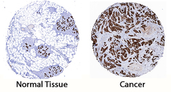
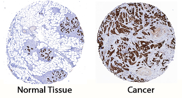
- Cytokeratin 19 in Human Breast Cancer Tissue. Cytokeratin 19 was detected in immersion fixed paraffin-embedded sections of human breast cancer tissue using Mouse Anti-Human Cytokeratin 19 Monoclonal Antibody (Catalog # MAB35061) at 5 µg/mL for 1 hour at room temperature followed by incubation with the Anti-Mouse IgG VisUCyte™ HRP Polymer Antibody (Catalog # VC001). Tissue was stained using DAB (brown) and counterstained with hematoxylin (blue). Specific staining was localized to cytoplasm. View our protocol for IHC Staining with VisUCyte HRP Polymer Detection Reagents.
- Submitted by
- R&D Systems (provider)
- Main image
- Experimental details
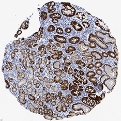
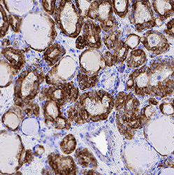
- Cytokeratin 19 in Human Gastric Cancer Tissue. Cytokeratin 19 was detected in immersion fixed paraffin-embedded sections of human gastric cancer tissue using Mouse Anti-Human Cytokeratin 19 Monoclonal Antibody (Catalog # MAB35061) at 0.5 µg/mL for 1 hour at room temperature followed by incubation with the Anti-Mouse IgG VisUCyte™ HRP Polymer Antibody (Catalog # VC001). Tissue was stained using DAB (brown) and counterstained with hematoxylin (blue). Specific staining was localized to cytoplasm in gastric glands. View our protocol for IHC Staining with VisUCyte HRP Polymer Detection Reagents.
- Submitted by
- R&D Systems (provider)
- Main image
- Experimental details
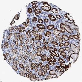
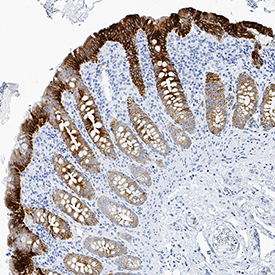
- Cytokeratin 19 in Human Papillary Thyroid Cancer Tissue. Cytokeratin 19 was detected in immersion fixed paraffin-embedded sections of human papillary thyroid cancer tissue using Mouse Anti-Human Cytokeratin 19 Monoclonal Antibody (Catalog # MAB35061) at 0.5 µg/mL for 1 hour at room temperature followed by incubation with the Anti-Mouse IgG VisUCyte™ HRP Polymer Antibody (Catalog # VC001). Tissue was stained using DAB (brown) and counterstained with hematoxylin (blue). Specific staining was localized to cytoplasm in epithelial cells. View our protocol for IHC Staining with VisUCyte HRP Polymer Detection Reagents.

- Submitted by
- R&D Systems (provider)
- Main image

- Experimental details
- Cytokeratin 19 in Human Lymphoma Tissue. Cytokeratin 19 was detected in immersion fixed paraffin-embedded sections of human lymphoma tissue using Mouse Anti-Human Cytokeratin 19 Monoclonal Antibody (Catalog # MAB35061) at 0.5 µg/mL for 1 hour at room temperature followed by incubation with the Anti-Mouse IgG VisUCyte™ HRP Polymer Antibody (Catalog # VC001). Tissue was stained using DAB (brown) and counterstained with hematoxylin (blue). Specific staining was localized to cell surfaces. View our protocol for IHC Staining with VisUCyte HRP Polymer Detection Reagents.
Supportive validation
- Submitted by
- R&D Systems (provider)
- Main image

- Experimental details
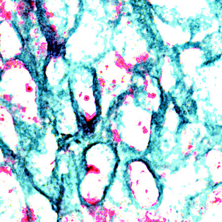
- Cytokeratin 19 in Human Breast Cancer Tissue Using Dual RNAscope® ISH and IHC. Cytokeratin 19 mRNA (red) and protein (green) was detected in formalin-fixed paraffin-embedded tissue sections of human breast cancer tissue probed with ACD RNAScope® Probe (Catalog # 310221) followed by immunohistochemistry using R&D Systems Mouse Anti-Human Cytokeratin 19 Monoclonal Antibody (Catalog # MAB35061) at 15ug/mL for 1 hour at room temperature followed by incubation with the Anti-Mouse IgG VisUCyte HRP Polymer Antibody (R&D Systems, Catalog # VC001). Tissue was stained using ACD RNAscope® 2.5 HD Duplex Detection Reagents (Catalog # 322500).