Explore
 Validate
 Learn
Learn Western blot
Antibody data
- Antibody Data
- Antigen structure
- References [7]
- Comments [0]
- Validations
- Western blot [1]
- Immunocytochemistry [2]
Submit
Validation data
Reference
Comment
Report error
- Product number
- 14-9898-82 - Provider product page

- Provider
- Invitrogen Antibodies
- Product name
- Cytokeratin 19 Monoclonal Antibody (BA17), eBioscience™
- Antibody type
- Monoclonal
- Antigen
- Other
- Description
- Description: This BA17 monoclonal antibody reacts with human cytokeratin 19 (CK19), a 44-kDa type I (acidic) intermediate filament protein that lacks the non-alpha-helical tail domain present in other keratins. Cytokeratins form the intracellular cytoskeletal network that maintains the integrity and stability of cells and tissues. Cytokeratin 19 is expressed in a wide variety of simple and stratified epithelial tissue. Moreover, cytokeratin 19 expression can be induced by vitamin A, SV40 transformation, and cancer. A soluble form of cytokeratin 19 generated by caspase 3 cleavage has also been found to be secreted by cancer cells, thus possibly indicating tumor metastasis. Cytokeratin 19 often exists as a heterodimer with cytokeratin 7, a type II keratin.
- Antibody clone number
- BA17
- Concentration
- 0.5 mg/mL
Submitted references Adjuvant gemcitabine therapy improves survival in a locally induced, R0-resectable model of metastatic intrahepatic cholangiocarcinoma.
Epidermal IL-15Rα acts as an endogenous antagonist of psoriasiform inflammation in mouse and man.
Expression of intercellular adhesion molecule 1 by hepatocellular carcinoma stem cells and circulating tumor cells.
Full-length cytokeratin-19 is released by human tumor cells: a potential role in metastatic progression of breast cancer.
The human keratins: biology and pathology.
Differential expression of keratin 19 in normal human epithelial tissues revealed by monospecific monoclonal antibodies.
Amino acid sequence and gene organization of cytokeratin no. 19, an exceptional tail-less intermediate filament protein.
Gürlevik E, Fleischmann-Mundt B, Armbrecht N, Longerich T, Woller N, Kloos A, Hoffmann D, Schambach A, Wirth TC, Manns MP, Zender L, Kubicka S, Kühnel F
Hepatology (Baltimore, Md.) 2013 Sep;58(3):1031-41
Hepatology (Baltimore, Md.) 2013 Sep;58(3):1031-41
Epidermal IL-15Rα acts as an endogenous antagonist of psoriasiform inflammation in mouse and man.
Bouchaud G, Gehrke S, Krieg C, Kolios A, Hafner J, Navarini AA, French LE, Boyman O
The Journal of experimental medicine 2013 Sep 23;210(10):2105-17
The Journal of experimental medicine 2013 Sep 23;210(10):2105-17
Expression of intercellular adhesion molecule 1 by hepatocellular carcinoma stem cells and circulating tumor cells.
Liu S, Li N, Yu X, Xiao X, Cheng K, Hu J, Wang J, Zhang D, Cheng S, Liu S
Gastroenterology 2013 May;144(5):1031-1041.e10
Gastroenterology 2013 May;144(5):1031-1041.e10
Full-length cytokeratin-19 is released by human tumor cells: a potential role in metastatic progression of breast cancer.
Alix-Panabières C, Vendrell JP, Slijper M, Pellé O, Barbotte E, Mercier G, Jacot W, Fabbro M, Pantel K
Breast cancer research : BCR 2009;11(3):R39
Breast cancer research : BCR 2009;11(3):R39
The human keratins: biology and pathology.
Moll R, Divo M, Langbein L
Histochemistry and cell biology 2008 Jun;129(6):705-33
Histochemistry and cell biology 2008 Jun;129(6):705-33
Differential expression of keratin 19 in normal human epithelial tissues revealed by monospecific monoclonal antibodies.
Bártek J, Bártková J, Taylor-Papadimitriou J, Rejthar A, Kovarík J, Lukás Z, Vojtĕsek B
The Histochemical journal 1986 Oct;18(10):565-75
The Histochemical journal 1986 Oct;18(10):565-75
Amino acid sequence and gene organization of cytokeratin no. 19, an exceptional tail-less intermediate filament protein.
Bader BL, Magin TM, Hatzfeld M, Franke WW
The EMBO journal 1986 Aug;5(8):1865-75
The EMBO journal 1986 Aug;5(8):1865-75
No comments: Submit comment
Supportive validation
- Submitted by
- Invitrogen Antibodies (provider)
- Main image
- Experimental details
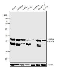
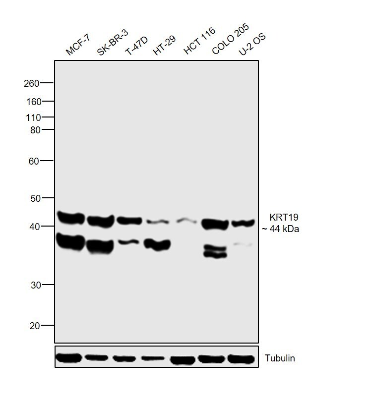
- Western blot was performed using Anti-Cytokeratin 19 Monoclonal Antibody (BA17), eBioscience™ (Product # 14-9898-82) and a 44 kDa band corresponding to KRT19 was observed across cell lines tested. An additional band around 38 kDa has been observed which is reported for Cytokeratin 19 expression. Membrane enriched extracts (30 µg lysate) of MCF-7 (Lane 1), SK-BR-3 (Lane 2), T-47D (Lane 3), HT-29 (Lane 4), HCT 116 (Lane 5), COLO 205 (Lane 6) and U-2 OS (Lane 7) were electrophoresed using NuPAGE™ 4-12% Bis-Tris Protein Gel (Product # NP0322BOX). Resolved proteins were then transferred onto a nitrocellulose membrane (Product # IB23001) by iBlot® 2 Dry Blotting System (Product # IB21001). The blot was probed with the primary antibody (1 ug/ml) and detected by chemiluminescence with Goat anti-Mouse IgG (H+L) Superclonal™ Recombinant Secondary Antibody, HRP (Product # A28177, 1:4000 dilution) using the iBright FL 1000 (Product # A32752). Chemiluminescent detection was performed using Novex® ECL Chemiluminescent Substrate Reagent Kit (Product # WP20005).
Supportive validation
- Submitted by
- Invitrogen Antibodies (provider)
- Main image

- Experimental details
- Immunocytochemistry of fixed and permeabilized MCF-7 cells using 10 µg/mL of Mouse IgG1 kappa Isotype Control (Product # 14-4714-82) (left) or 10 µg/mL of Anti-Cytokeratin 19 Purified (right) followed by Anti-Mouse TRITC.