Explore
 Validate
 Learn
Learn Western blot
 Immunohistochemistry
Antibody data
- Antibody Data
- Antigen structure
- References [2]
- Comments [0]
- Validations
- Immunohistochemistry [1]
Submit
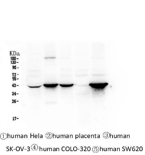
Validation data
Reference
Comment
Report error
- Product number
- M02101-2 - Provider product page

- Provider
- Boster Biological Technology
- Product name
- Anti-Cytokeratin 19 KRT19 Antibody Picoband™ (monoclonal, 3D4)
- Antibody type
- Monoclonal
- Description
- Mouse IgG monoclonal antibody for Cytokeratin 19 detection. Tested with WB, IHC-P, IF in Human.
- Reactivity
- Human
- Host
- Mouse
- Isotype
- IgG
- Antibody clone number
- 3D4
- Vial size
- 100μg/vial
- Concentration
- 0.5-1mg/ml, actual concentration vary by lot. Use suggested dilution ratio to decide dilution procedure.
- Storage
- At -20°C for one year. After reconstitution, at 4°C for one month. It can also be aliquoted and stored frozen at -20°C for a longer time. Avoid repeated freezing and thawing.
- Handling
- Add 0.2ml of distilled water will yield a concentration of 500ug/ml.
Submitted references Necroptosis-Related Prognostic Model for Pancreatic Carcinoma Reveals Its Invasion and Metastasis Potential through Hybrid EMT and Immune Escape.
Establishment and evaluation of the goose embryo epithelial (GEE) cell line as a new model for propagation of avian viruses.
Liu H, Li Z, Zhang L, Zhang M, Liu S, Wang J, Yang C, Peng Q, Du C, Jiang N
Biomedicines 2023 Jun 16;11(6)
Biomedicines 2023 Jun 16;11(6)
Establishment and evaluation of the goose embryo epithelial (GEE) cell line as a new model for propagation of avian viruses.
Wang W, Said A, Wang B, Qu G, Xu Q, Liu B, Shen Z
PloS one 2018;13(3):e0193876
PloS one 2018;13(3):e0193876
No comments: Submit comment
Supportive validation
- Submitted by
- Boster Biological Technology (provider)
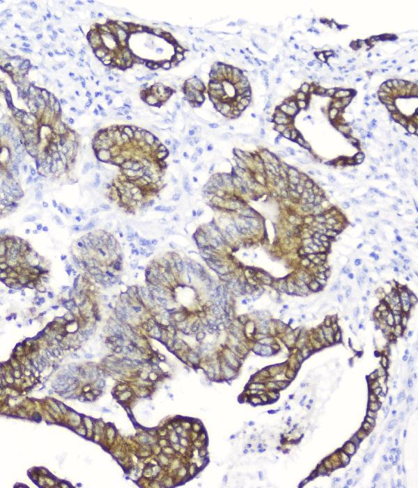
- Main image
- Experimental details
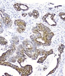
- IHC analysis of Cytokeratin 19 using anti-Cytokeratin 19 antibody (M02101-2). Cytokeratin 19 was detected in paraffin-embedded section of human intestinal cancer tissue. Heat mediated antigen retrieval was performed in citrate buffer (pH6, epitope retrieval solution) for 20 mins. The tissue section was blocked with 10% goat serum. The tissue section was then incubated with 2μg/ml mouse anti-Cytokeratin 19 Antibody (M02101-2) overnight at 4°C. Biotinylated goat anti-mouse IgG was used as secondary antibody and incubated for 30 minutes at 37°C. The tissue section was developed using Strepavidin-Biotin-Complex (SABC)(Catalog # SA1021) with DAB as the chromogen.
- Additional image