Explore
 Validate
 Learn
Learn Western blot
 Immunocytochemistry
Antibody data
- Antibody Data
- Antigen structure
- References [53]
- Comments [0]
- Validations
- Immunocytochemistry [2]
- Immunohistochemistry [1]
- Flow cytometry [1]
- Other assay [11]
Submit
Validation data
Reference
Comment
Report error
- Product number
- MA5-12663 - Provider product page

- Provider
- Invitrogen Antibodies
- Product name
- Cytokeratin 19 Monoclonal Antibody (A53-B/A2.26 (Ks19.1))
- Antibody type
- Monoclonal
- Antigen
- Other
- Description
- MA5-12663 targets Cytokeratin 19 in IF, IHC (P), and WB applications and shows reactivity with Human samples. The MA5-12663 immunogen is human breast cancer MCF-7 cells.
- Reactivity
- Human
- Host
- Mouse
- Isotype
- IgG
- Antibody clone number
- A53-B/A2.26 (Ks19.1)
- Vial size
- 500 μL
- Concentration
- 0.2 mg/mL
- Storage
- 4°C
Submitted references Downregulation of MMP-9 Enhances the Anti-Migratory Effect of Cyclophosphamide in MDA-MB-231 and MCF-7 Breast Cancer Cell Lines.
Immunohistochemical evaluation of hepatic progenitor cells in different types of feline liver diseases.
Expression of keratins 8, 18, and 19 in epithelia of atrophic oral lichen planus.
Propagation of functional estrogen receptor positive normal human breast cells in 3D cultures.
Patient-derived pancreas-on-a-chip to model cystic fibrosis-related disorders.
Integrated Molecular Characterization of Uterine Carcinosarcoma.
The Hippo kinases LATS1 and 2 control human breast cell fate via crosstalk with ERα.
CD56, HBME-1 and cytokeratin 19 expressions in papillary thyroid carcinoma and nodular thyroid lesions.
Thyroid Paraganglioma Diagnosed by Fine-Needle Aspiration Biopsy, Correlated With Histopathological Findings: Report of a Case.
Galectin-3 and HBME-1 improve the accuracy of core biopsy in indeterminate thyroid nodules.
Clinicopathological characteristics of kidney mucinous tubular and spindle cell carcinoma.
Potential Cross-Talk between Alternative and Classical NF-κB Pathways in Prostate Cancer Tissues as Measured by a Multi-Staining Immunofluorescence Co-Localization Assay.
Novel high-grade serous epithelial ovarian cancer cell lines that reflect the molecular diversity of both the sporadic and hereditary disease.
Novel high-grade serous epithelial ovarian cancer cell lines that reflect the molecular diversity of both the sporadic and hereditary disease.
Keratin 19 protein expression is an independent predictor of survival in human hepatocellular carcinoma.
Diagnostic criteria of well differentiated thyroid tumor of uncertain malignant potential; a histomorphological and immunohistochemical appraisal.
High mammographic density is associated with an increase in stromal collagen and immune cells within the mammary epithelium.
Modelling breast cancer requires identification and correction of a critical cell lineage-dependent transduction bias.
Keratinocyte growth factor-2 and autologous serum potentiate the regenerative effect of mesenchymal stem cells in cornea damage in rats.
Molecular features of follicular variant papillary carcinoma of thyroid: comparison of areas with or without classical nuclear features.
Thyroid-like papillary adenocarcinoma of the nasopharynx with focal thyroglobulin expression.
A Case of Multifocal Papillary Thyroid Carcinoma Consisting of One Encapsulated Follicular Variant with BRAF K601E Mutation and Three Conventional Types with BRAF V600E Mutation.
Establishment and characterization of a telomerase immortalized human gingival epithelial cell line.
A renewable tissue resource of phenotypically stable, biologically and ethnically diverse, patient-derived human breast cancer xenograft models.
Establishment and characterization of novel cell lines from sinonasal undifferentiated carcinoma.
Gremlin in the pathogenesis of hepatocellular carcinoma complicating chronic hepatitis C: an immunohistochemical and PCR study of human liver biopsies.
Primary pure squamous cell carcinoma of the thyroid: report and histogenic consideration of a case involving a BRAF mutation.
Derivation and characterization of matched cell lines from primary and recurrent serous ovarian cancer.
Mucinous tubular and spindle cell carcinoma of kidney and problems in diagnosis.
Comparison of a poly-L-lactide-co-ε-caprolactone and human amniotic membrane for urothelium tissue engineering applications.
Tumor-in-tumor of the thyroid with basaloid differentiation: a lesion with a solid cell nest neoplastic component?
In vitro constitution and in vivo implantation of engineered skin constructs with sweat glands.
Human embryonic stem cell-derived keratinocytes exhibit an epidermal transcription program and undergo epithelial morphogenesis in engineered tissue constructs.
Tissue proteomics of the human mammary gland: towards an abridged definition of the molecular phenotypes underlying epithelial normalcy.
Assessment of the tumorigenesis and drug susceptibility of three new canine mammary tumor cell lines.
An intraductal human-in-mouse transplantation model mimics the subtypes of ductal carcinoma in situ.
Antigen expression of human eccrine sweat glands.
Nucleostemin as a possible progenitor marker of corneal epithelial cells.
Absence of the BRAF mutation in HBME1+ and CK19+ atypical cell clusters in Hashimoto thyroiditis: supportive evidence against preneoplastic change.
15-prostaglandin dehydrogenase expression alone or in combination with ACSM1 defines a subgroup of the apocrine molecular subtype of breast carcinoma.
Isolation and identification of stem cells from adult cashmere goat skin.
Expression of estrogen-metabolizing enzymes and estrogen receptors in cholelithiasis gallbladder.
An immunohistochemical profile of giant cell carcinoma of the larynx.
Mammary gland development in early pubertal female macaques.
Characterization of breast precancerous lesions and myoepithelial hyperplasia in sclerosing adenosis with apocrine metaplasia.
Isolation and characterization of putative epidermal stem cells derived from Cashmere goat fetus.
Identification of a subset of breast carcinomas characterized by expression of cytokeratin 15: relationship between CK15+ progenitor/amplified cells and pre-malignant lesions and invasive disease.
Immunohistochemical markers in diagnosis of papillary thyroid carcinoma: Utility of HBME1 combined with CK19 immunostaining.
Mesenchymal hamartoma of the liver in adulthood: immunohistochemical profiles, clinical and histopathological features in two patients.
Down-regulation of the tumor suppressor protein 14-3-3sigma is a sporadic event in cancer of the breast.
Cytokeratin expression in lichen amyloidosus and macular amyloidosis.
Subcellular localization of the ABCG2 transporter in normal and malignant human gallbladder epithelium.
Isolation, immortalization, and characterization of a human breast epithelial cell line with stem cell properties.
Izdebska M, Zielińska W, Krajewski A, Hałas-Wiśniewska M, Mikołajczyk K, Gagat M, Grzanka A
International journal of molecular sciences 2021 Nov 26;22(23)
International journal of molecular sciences 2021 Nov 26;22(23)
Immunohistochemical evaluation of hepatic progenitor cells in different types of feline liver diseases.
Abou Monsef Y, Kutsal O
The Journal of veterinary medical science 2021 Apr 9;83(4):613-621
The Journal of veterinary medical science 2021 Apr 9;83(4):613-621
Expression of keratins 8, 18, and 19 in epithelia of atrophic oral lichen planus.
Schreurs O, Karatsaidis A, Balta MG, Grung B, Hals EKB, Schenck K
European journal of oral sciences 2020 Feb;128(1):7-17
European journal of oral sciences 2020 Feb;128(1):7-17
Propagation of functional estrogen receptor positive normal human breast cells in 3D cultures.
Meng P, Vaapil M, Tagmount A, Loguinov A, Vulpe C, Yaswen P
Breast cancer research and treatment 2019 Jul;176(1):131-140
Breast cancer research and treatment 2019 Jul;176(1):131-140
Patient-derived pancreas-on-a-chip to model cystic fibrosis-related disorders.
Shik Mun K, Arora K, Huang Y, Yang F, Yarlagadda S, Ramananda Y, Abu-El-Haija M, Palermo JJ, Appakalai BN, Nathan JD, Naren AP
Nature communications 2019 Jul 16;10(1):3124
Nature communications 2019 Jul 16;10(1):3124
Integrated Molecular Characterization of Uterine Carcinosarcoma.
Cherniack AD, Shen H, Walter V, Stewart C, Murray BA, Bowlby R, Hu X, Ling S, Soslow RA, Broaddus RR, Zuna RE, Robertson G, Laird PW, Kucherlapati R, Mills GB, Cancer Genome Atlas Research Network, Weinstein JN, Zhang J, Akbani R, Levine DA
Cancer cell 2017 Mar 13;31(3):411-423
Cancer cell 2017 Mar 13;31(3):411-423
The Hippo kinases LATS1 and 2 control human breast cell fate via crosstalk with ERα.
Britschgi A, Duss S, Kim S, Couto JP, Brinkhaus H, Koren S, De Silva D, Mertz KD, Kaup D, Varga Z, Voshol H, Vissieres A, Leroy C, Roloff T, Stadler MB, Scheel CH, Miraglia LJ, Orth AP, Bonamy GM, Reddy VA, Bentires-Alj M
Nature 2017 Jan 26;541(7638):541-545
Nature 2017 Jan 26;541(7638):541-545
CD56, HBME-1 and cytokeratin 19 expressions in papillary thyroid carcinoma and nodular thyroid lesions.
Erdogan-Durmus S, Ozcan D, Yarikkaya E, Kurt A, Arslan A
Journal of research in medical sciences : the official journal of Isfahan University of Medical Sciences 2016;21:49
Journal of research in medical sciences : the official journal of Isfahan University of Medical Sciences 2016;21:49
Thyroid Paraganglioma Diagnosed by Fine-Needle Aspiration Biopsy, Correlated With Histopathological Findings: Report of a Case.
Çetin Ş, Kir G, Yilmaz M
Diagnostic cytopathology 2016 Jul;44(7):643-7
Diagnostic cytopathology 2016 Jul;44(7):643-7
Galectin-3 and HBME-1 improve the accuracy of core biopsy in indeterminate thyroid nodules.
Trimboli P, Guidobaldi L, Amendola S, Nasrollah N, Romanelli F, Attanasio D, Ramacciato G, Saggiorato E, Valabrega S, Crescenzi A
Endocrine 2016 Apr;52(1):39-45
Endocrine 2016 Apr;52(1):39-45
Clinicopathological characteristics of kidney mucinous tubular and spindle cell carcinoma.
Chen Q, Gu Y, Liu B
International journal of clinical and experimental pathology 2015;8(1):1007-12
International journal of clinical and experimental pathology 2015;8(1):1007-12
Potential Cross-Talk between Alternative and Classical NF-κB Pathways in Prostate Cancer Tissues as Measured by a Multi-Staining Immunofluorescence Co-Localization Assay.
Labouba I, Le Page C, Communal L, Kristessen T, You X, Péant B, Barrès V, Gannon PO, Mes-Masson AM, Saad F
PloS one 2015;10(7):e0131024
PloS one 2015;10(7):e0131024
Novel high-grade serous epithelial ovarian cancer cell lines that reflect the molecular diversity of both the sporadic and hereditary disease.
Fleury H, Communal L, Carmona E, Portelance L, Arcand SL, Rahimi K, Tonin PN, Provencher D, Mes-Masson AM
Genes & cancer 2015 Sep;6(9-10):378-398
Genes & cancer 2015 Sep;6(9-10):378-398
Keratin 19 protein expression is an independent predictor of survival in human hepatocellular carcinoma.
Fatourou E, Koskinas J, Karandrea D, Palaiologou M, Syminelaki T, Karanikolas M, Felekouras E, Antoniou E, Manesis EK, Delladetsima J, Tiniakos D
European journal of gastroenterology & hepatology 2015 Sep;27(9):1094-102
European journal of gastroenterology & hepatology 2015 Sep;27(9):1094-102
Diagnostic criteria of well differentiated thyroid tumor of uncertain malignant potential; a histomorphological and immunohistochemical appraisal.
Yassin Fel-Z
Journal of the Egyptian National Cancer Institute 2015 Jun;27(2):59-67
Journal of the Egyptian National Cancer Institute 2015 Jun;27(2):59-67
High mammographic density is associated with an increase in stromal collagen and immune cells within the mammary epithelium.
Huo CW, Chew G, Hill P, Huang D, Ingman W, Hodson L, Brown KA, Magenau A, Allam AH, McGhee E, Timpson P, Henderson MA, Thompson EW, Britt K
Breast cancer research : BCR 2015 Jun 4;17(1):79
Breast cancer research : BCR 2015 Jun 4;17(1):79
Modelling breast cancer requires identification and correction of a critical cell lineage-dependent transduction bias.
Hines WC, Yaswen P, Bissell MJ
Nature communications 2015 Apr 21;6:6927
Nature communications 2015 Apr 21;6:6927
Keratinocyte growth factor-2 and autologous serum potentiate the regenerative effect of mesenchymal stem cells in cornea damage in rats.
Pınarlı FA, Okten G, Beden U, Fışgın T, Kefeli M, Kara N, Duru F, Tomak L
International journal of ophthalmology 2014;7(2):211-9
International journal of ophthalmology 2014;7(2):211-9
Molecular features of follicular variant papillary carcinoma of thyroid: comparison of areas with or without classical nuclear features.
Guney G, Tezel GG, Kosemehmetoglu K, Yilmaz E, Balci S, Ersoy R, Cakir B, Guler G
Endocrine pathology 2014 Sep;25(3):241-7
Endocrine pathology 2014 Sep;25(3):241-7
Thyroid-like papillary adenocarcinoma of the nasopharynx with focal thyroglobulin expression.
Ozer S, Kayahan B, Cabbarzade C, Bugdayci M, Kosemehmetoglu K, Yucel OT
Pathology 2013 Oct;45(6):622-4
Pathology 2013 Oct;45(6):622-4
A Case of Multifocal Papillary Thyroid Carcinoma Consisting of One Encapsulated Follicular Variant with BRAF K601E Mutation and Three Conventional Types with BRAF V600E Mutation.
Kim WY, Ko YS, Hwang TS, Han HS, Lim SD, Kim WS, Oh SY
Korean journal of pathology 2013 Jun;47(3):293-8
Korean journal of pathology 2013 Jun;47(3):293-8
Establishment and characterization of a telomerase immortalized human gingival epithelial cell line.
Moffatt-Jauregui CE, Robinson B, de Moya AV, Brockman RD, Roman AV, Cash MN, Culp DJ, Lamont RJ
Journal of periodontal research 2013 Dec;48(6):713-21
Journal of periodontal research 2013 Dec;48(6):713-21
A renewable tissue resource of phenotypically stable, biologically and ethnically diverse, patient-derived human breast cancer xenograft models.
Zhang X, Claerhout S, Prat A, Dobrolecki LE, Petrovic I, Lai Q, Landis MD, Wiechmann L, Schiff R, Giuliano M, Wong H, Fuqua SW, Contreras A, Gutierrez C, Huang J, Mao S, Pavlick AC, Froehlich AM, Wu MF, Tsimelzon A, Hilsenbeck SG, Chen ES, Zuloaga P, Shaw CA, Rimawi MF, Perou CM, Mills GB, Chang JC, Lewis MT
Cancer research 2013 Aug 1;73(15):4885-97
Cancer research 2013 Aug 1;73(15):4885-97
Establishment and characterization of novel cell lines from sinonasal undifferentiated carcinoma.
Takahashi Y, Kupferman ME, Bell D, Jiffar T, Lee JG, Xie TX, Li NW, Zhao M, Frederick MJ, Gelbard A, Myers JN, Hanna EY
Clinical cancer research : an official journal of the American Association for Cancer Research 2012 Nov 15;18(22):6178-87
Clinical cancer research : an official journal of the American Association for Cancer Research 2012 Nov 15;18(22):6178-87
Gremlin in the pathogenesis of hepatocellular carcinoma complicating chronic hepatitis C: an immunohistochemical and PCR study of human liver biopsies.
Guimei M, Baddour N, Elkaffash D, Abdou L, Taher Y
BMC research notes 2012 Jul 29;5:390
BMC research notes 2012 Jul 29;5:390
Primary pure squamous cell carcinoma of the thyroid: report and histogenic consideration of a case involving a BRAF mutation.
Ko YS, Hwang TS, Han HS, Lim SD, Kim WS, Oh SY
Pathology international 2012 Jan;62(1):43-8
Pathology international 2012 Jan;62(1):43-8
Derivation and characterization of matched cell lines from primary and recurrent serous ovarian cancer.
Létourneau IJ, Quinn MC, Wang LL, Portelance L, Caceres KY, Cyr L, Delvoye N, Meunier L, de Ladurantaye M, Shen Z, Arcand SL, Tonin PN, Provencher DM, Mes-Masson AM
BMC cancer 2012 Aug 29;12:379
BMC cancer 2012 Aug 29;12:379
Mucinous tubular and spindle cell carcinoma of kidney and problems in diagnosis.
Sarsik B, Sımşır A, Karaarslan S, Sen S
Turk patoloji dergisi 2011 May;27(2):116-26
Turk patoloji dergisi 2011 May;27(2):116-26
Comparison of a poly-L-lactide-co-ε-caprolactone and human amniotic membrane for urothelium tissue engineering applications.
Sartoneva R, Haimi S, Miettinen S, Mannerström B, Haaparanta AM, Sándor GK, Kellomäki M, Suuronen R, Lahdes-Vasama T
Journal of the Royal Society, Interface 2011 May 6;8(58):671-7
Journal of the Royal Society, Interface 2011 May 6;8(58):671-7
Tumor-in-tumor of the thyroid with basaloid differentiation: a lesion with a solid cell nest neoplastic component?
Eloy C, Vinagre J, Cameselle-Teijeiro J, Paiva ME, Soares P, Sobrinho-Simões M
International journal of surgical pathology 2011 Apr;19(2):276-80
International journal of surgical pathology 2011 Apr;19(2):276-80
In vitro constitution and in vivo implantation of engineered skin constructs with sweat glands.
Huang S, Xu Y, Wu C, Sha D, Fu X
Biomaterials 2010 Jul;31(21):5520-5
Biomaterials 2010 Jul;31(21):5520-5
Human embryonic stem cell-derived keratinocytes exhibit an epidermal transcription program and undergo epithelial morphogenesis in engineered tissue constructs.
Metallo CM, Azarin SM, Moses LE, Ji L, de Pablo JJ, Palecek SP
Tissue engineering. Part A 2010 Jan;16(1):213-23
Tissue engineering. Part A 2010 Jan;16(1):213-23
Tissue proteomics of the human mammary gland: towards an abridged definition of the molecular phenotypes underlying epithelial normalcy.
Moreira JM, Cabezón T, Gromova I, Gromov P, Timmermans-Wielenga V, Machado I, Llombart-Bosch A, Kroman N, Rank F, Celis JE
Molecular oncology 2010 Dec;4(6):539-61
Molecular oncology 2010 Dec;4(6):539-61
Assessment of the tumorigenesis and drug susceptibility of three new canine mammary tumor cell lines.
Chang CY, Chiou PP, Chen WJ, Li YH, Yiu JC, Cheng YH, Chen SD, Lin CT, Lai YS
Research in veterinary science 2010 Apr;88(2):285-93
Research in veterinary science 2010 Apr;88(2):285-93
An intraductal human-in-mouse transplantation model mimics the subtypes of ductal carcinoma in situ.
Behbod F, Kittrell FS, LaMarca H, Edwards D, Kerbawy S, Heestand JC, Young E, Mukhopadhyay P, Yeh HW, Allred DC, Hu M, Polyak K, Rosen JM, Medina D
Breast cancer research : BCR 2009;11(5):R66
Breast cancer research : BCR 2009;11(5):R66
Antigen expression of human eccrine sweat glands.
Li HH, Zhou G, Fu XB, Zhang L
Journal of cutaneous pathology 2009 Mar;36(3):318-24
Journal of cutaneous pathology 2009 Mar;36(3):318-24
Nucleostemin as a possible progenitor marker of corneal epithelial cells.
Kawashima M, Kawakita T, Yoshida S, Shimmura S, Tsubota K
Molecular vision 2009 Jun 10;15:1162-8
Molecular vision 2009 Jun 10;15:1162-8
Absence of the BRAF mutation in HBME1+ and CK19+ atypical cell clusters in Hashimoto thyroiditis: supportive evidence against preneoplastic change.
Nasr MR, Mukhopadhyay S, Zhang S, Katzenstein AL
American journal of clinical pathology 2009 Dec;132(6):906-12
American journal of clinical pathology 2009 Dec;132(6):906-12
15-prostaglandin dehydrogenase expression alone or in combination with ACSM1 defines a subgroup of the apocrine molecular subtype of breast carcinoma.
Celis JE, Gromov P, Cabezón T, Moreira JM, Friis E, Jirström K, Llombart-Bosch A, Timmermans-Wielenga V, Rank F, Gromova I
Molecular & cellular proteomics : MCP 2008 Oct;7(10):1795-809
Molecular & cellular proteomics : MCP 2008 Oct;7(10):1795-809
Isolation and identification of stem cells from adult cashmere goat skin.
Liu Y, Zhou H, Gao F
International journal of dermatology 2008 Jun;47(6):551-6
International journal of dermatology 2008 Jun;47(6):551-6
Expression of estrogen-metabolizing enzymes and estrogen receptors in cholelithiasis gallbladder.
Svoboda M, Sellner F, Ekmekcioglu C, Klimpfinger M, Jaeger W, Thalhammer T
Biomedicine & pharmacotherapy = Biomedecine & pharmacotherapie 2008 Dec;62(10):690-6
Biomedicine & pharmacotherapy = Biomedecine & pharmacotherapie 2008 Dec;62(10):690-6
An immunohistochemical profile of giant cell carcinoma of the larynx.
Gurbuz Y, Kose N, Aydin O, Ozturk M
Auris, nasus, larynx 2007 Sep;34(3):413-6
Auris, nasus, larynx 2007 Sep;34(3):413-6
Mammary gland development in early pubertal female macaques.
Wood CE, Hester JM, Cline JM
Toxicologic pathology 2007 Oct;35(6):795-805
Toxicologic pathology 2007 Oct;35(6):795-805
Characterization of breast precancerous lesions and myoepithelial hyperplasia in sclerosing adenosis with apocrine metaplasia.
Celis JE, Moreira JM, Gromova I, Cabezón T, Gromov P, Shen T, Timmermans V, Rank F
Molecular oncology 2007 Jun;1(1):97-119
Molecular oncology 2007 Jun;1(1):97-119
Isolation and characterization of putative epidermal stem cells derived from Cashmere goat fetus.
Islam MS, Zhou H
European journal of dermatology : EJD 2007 Jul-Aug;17(4):302-8
European journal of dermatology : EJD 2007 Jul-Aug;17(4):302-8
Identification of a subset of breast carcinomas characterized by expression of cytokeratin 15: relationship between CK15+ progenitor/amplified cells and pre-malignant lesions and invasive disease.
Celis JE, Gromova I, Cabezón T, Gromov P, Shen T, Timmermans-Wielenga V, Rank F, Moreira JM
Molecular oncology 2007 Dec;1(3):321-49
Molecular oncology 2007 Dec;1(3):321-49
Immunohistochemical markers in diagnosis of papillary thyroid carcinoma: Utility of HBME1 combined with CK19 immunostaining.
Nasr MR, Mukhopadhyay S, Zhang S, Katzenstein AL
Modern pathology : an official journal of the United States and Canadian Academy of Pathology, Inc 2006 Dec;19(12):1631-7
Modern pathology : an official journal of the United States and Canadian Academy of Pathology, Inc 2006 Dec;19(12):1631-7
Mesenchymal hamartoma of the liver in adulthood: immunohistochemical profiles, clinical and histopathological features in two patients.
Yesim G, Gupse T, Zafer U, Ahmet A
Journal of hepato-biliary-pancreatic surgery 2005;12(6):502-7
Journal of hepato-biliary-pancreatic surgery 2005;12(6):502-7
Down-regulation of the tumor suppressor protein 14-3-3sigma is a sporadic event in cancer of the breast.
Moreira JM, Ohlsson G, Rank FE, Celis JE
Molecular & cellular proteomics : MCP 2005 Apr;4(4):555-69
Molecular & cellular proteomics : MCP 2005 Apr;4(4):555-69
Cytokeratin expression in lichen amyloidosus and macular amyloidosis.
Apaydin R, Gürbüz Y, Bayramgürler D, Müezzinoglu B, Bilen N
Journal of the European Academy of Dermatology and Venereology : JEADV 2004 May;18(3):305-9
Journal of the European Academy of Dermatology and Venereology : JEADV 2004 May;18(3):305-9
Subcellular localization of the ABCG2 transporter in normal and malignant human gallbladder epithelium.
Aust S, Obrist P, Jaeger W, Klimpfinger M, Tucek G, Wrba F, Penner E, Thalhammer T
Laboratory investigation; a journal of technical methods and pathology 2004 Aug;84(8):1024-36
Laboratory investigation; a journal of technical methods and pathology 2004 Aug;84(8):1024-36
Isolation, immortalization, and characterization of a human breast epithelial cell line with stem cell properties.
Gudjonsson T, Villadsen R, Nielsen HL, Rønnov-Jessen L, Bissell MJ, Petersen OW
Genes & development 2002 Mar 15;16(6):693-706
Genes & development 2002 Mar 15;16(6):693-706
No comments: Submit comment
Supportive validation
- Submitted by
- Invitrogen Antibodies (provider)
- Main image
- Experimental details
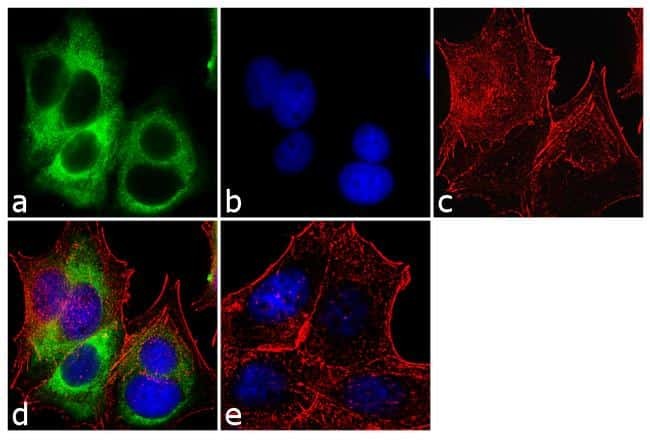
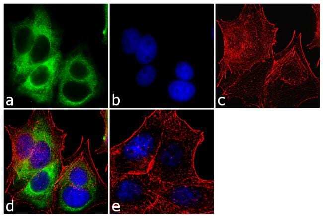
- Immunofluorescent analysis of Cytokeratin 19 was performed using 70% confluent log phase MCF-7 cells. The cells were fixed with 4% paraformaldehyde for 10 minutes, permeabilized with 0.1% Triton™ X-100 for 10 minutes, and blocked with 1% BSA for 1 hour at room temperature. The cells were labeled with Cytokeratin 19 (A53-B/A2.26 (Ks19.1)) Mouse Monoclonal Antibody (Product # MA5-12663) at 2 µg/mL in 0.1% BSA and incubated for 3 hours at room temperature and then labeled with Goat anti-Mouse IgG (H+L) Superclonal™ Secondary Antibody, Alexa Fluor® 488 conjugate (Product # A28175) a dilution of 1:2000 for 45 minutes at room temperature (Panel a: green). Nuclei (Panel b: blue) were stained with SlowFade® Gold Antifade Mountant with DAPI (Product # S36938). F-actin (Panel c: red) was stained with Alexa Fluor® 555 Rhodamine Phalloidin (Product # R415, 1:300). Panel d represents the merged image showing cytoplasmic localization. Panel e shows the no primary antibody control. The images were captured at 60X magnification.
- Submitted by
- Invitrogen Antibodies (provider)
- Main image
- Experimental details
- Immunofluorescent analysis of Cytokeratin 19 was performed using 70% confluent log phase MCF-7 cells. The cells were fixed with 4% paraformaldehyde for 10 minutes, permeabilized with 0.1% Triton™ X-100 for 10 minutes, and blocked with 1% BSA for 1 hour at room temperature. The cells were labeled with Cytokeratin 19 (A53-B/A2.26 (Ks19.1)) Mouse Monoclonal Antibody (Product # MA5-12663) at 2 µg/mL in 0.1% BSA and incubated for 3 hours at room temperature and then labeled with Goat anti-Mouse IgG (H+L) Superclonal™ Secondary Antibody, Alexa Fluor® 488 conjugate (Product # A28175) a dilution of 1:2000 for 45 minutes at room temperature (Panel a: green). Nuclei (Panel b: blue) were stained with SlowFade® Gold Antifade Mountant with DAPI (Product # S36938). F-actin (Panel c: red) was stained with Alexa Fluor® 555 Rhodamine Phalloidin (Product # R415, 1:300). Panel d represents the merged image showing cytoplasmic localization. Panel e shows the no primary antibody control. The images were captured at 60X magnification.
Supportive validation
- Submitted by
- Invitrogen Antibodies (provider)
- Main image
- Experimental details
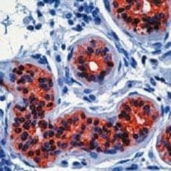
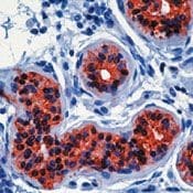
- Formalin-fixed, paraffin-embedded human skin stained with Keratin 19 antibody using peroxidase-conjugate and AEC chromogen. Note cytoplasmic staining of epithelial cells in the sweat glands.
Supportive validation
- Submitted by
- Invitrogen Antibodies (provider)
- Main image
- Experimental details
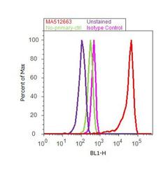
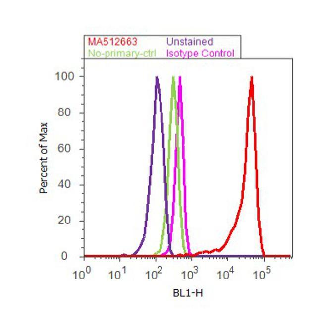
- Flow cytometry analysis of Cytokeratin 19 was done on MCF7 cells. Cells were fixed with 70% ethanol for 10 minutes, permeabilized with 0.25% Triton™ X-100 for 20 minutes, and blocked with 5% BSA for 30 minutes at room temperature. Cells were labeled with Cytokeratin 19 Mouse Monoclonal Antibody (MA5-12663, red histogram) or with mouse isotype control (pink histogram) at 3-5 ug/million cells in 2.5% BSA. After incubation at room temperature for 2 hours, the cells were labeled with Alexa Fluor® 488 Rabbit Anti-Mouse Secondary Antibody (A11059) at a dilution of 1:400 for 30 minutes at room temperature. The representative 10, 000 cells were acquired and analyzed for each sample using an Attune® Acoustic Focusing Cytometer. The purple histogram represents unstained control cells and the green histogram represents no-primary-antibody control.
Supportive validation
- Submitted by
- Invitrogen Antibodies (provider)
- Main image
- Experimental details
- NULL
- Submitted by
- Invitrogen Antibodies (provider)
- Main image
- Experimental details
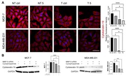
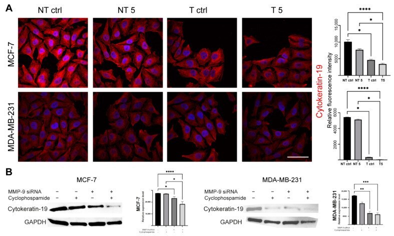
- Figure 8 The effect of MMP-9 downregulation on Cytokeratin 19 (CK19) level. NT--non-transfected cells, T--cells transfected with MMP-9 siRNA, NT5 and T5 non-transfected and transfected cells treated with 5 mM cyclophosphamide for 24 h. ( A ) Immunofluorescent imaging of Cytokeratin 19 (red), and nuclei (blue), and cytometric relative fluorescence intensity of Cytokeratin 19, Bar = 50 mum. ( B ) The Western blot analysis of Cytokeratin 19 and densitometric relative measurement of the Western blot experiments. Statistically significant differences were marked with '*' p
- Submitted by
- Invitrogen Antibodies (provider)
- Main image
- Experimental details
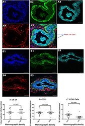
- Fig. 7 Cytokeratin (CK)-19, CK-14 and vimentin triple immunofluorescence staining. High mammographic density (HMD) ( a ) and low mammographic density (LMD) ( b ) tissue samples were stained with 4',6-diamidino-2-phenylindole ( a 1 and b 1 ), CK-14 ( a 2 and b 2 ) CK-19 ( a 3 and b 3 ) and vimentin ( a 4 and b 4 ) and assessed as composite images ( a 5 and b 5 ). Arrows indicate cells inside the epithelial layer that stained negative for CK-19 or CK-14, but positive for vimentin. ( c ) Using the point-counting method, the percentages of CK-14 basal epithelial cells and CK-19 luminal epithelial cells were determined, as were the number of vimentin-expressing CK - cells per whole glandular area of the tissue sections. The results for HMD and LMD paired samples were analysed using the nonparametric Wilcoxon matched-pairs signed-rank test. VPCKN vimentin-positive, cytokeratin-negative. Scale bar = 10 mum
- Submitted by
- Invitrogen Antibodies (provider)
- Main image
- Experimental details
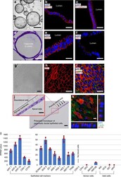
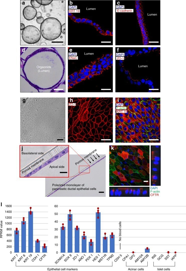
- Fig. 2 Characterization of pancreatic ductal epithelial cells. ( a - f , l : Characterization of organoids). a Characterization of pancreatic ductal organoids using epithelial cell markers, b cytokeratin 19 (KRT 19, c E-cadherin, e sodium transport channel (ENaC), and f ZO-1. d Hematoxylin and eosin (H&E) image shows the orientation of pancreatic ductal epithelial cells in spheroid of the organoid. ( g - k : Characterization of monolayer of pancreatic ductal epithelial cells (PDECs)). g Phase contrast and j H&E images show monolayer of PDECs formed from the organoids. Monolayer of PDECs showed positivity for tight junction h ZO-1, i F-actin and KRT 19, and k cystic fibrosis transmembrane conductance regulator (CFTR). l RNA-sequencing data was obtained from the pancreatic ductal organoids and verified the PDEC origin ( n = 4 sample preparation from the same patient). Data are mean +- SD. Scale bars: 10 um ( k ), 20 um ( b , c , e , f , h - j enlarge), 50 um ( d , g , j ), and 500 um ( a )
- Submitted by
- Invitrogen Antibodies (provider)
- Main image

- Experimental details
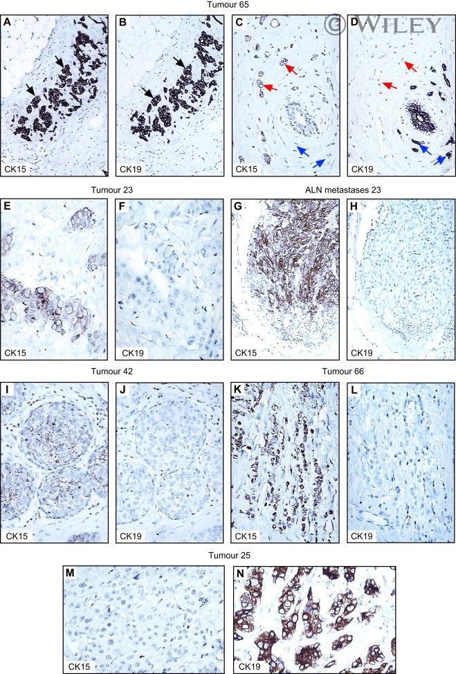
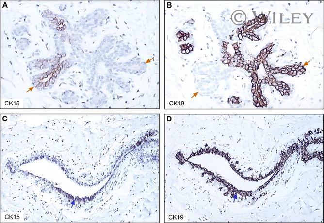
- IHC pictures of serial paraffin-embedded sections of tumours and ALN metastases stained with antibodies against CK15 (monoclonal) and CK19. Black arrows in (A) and (B) indicate cells that express both CK15 and CK19. Red arrows in (C) and (D) indicate cells that express only CK15, while blue arrows indicate cells that express only CK19.
- Submitted by
- Invitrogen Antibodies (provider)
- Main image
- Experimental details
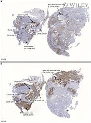
- IHC staining of serial paraffin-embedded sections of tumour 71 stained with antibodies against (A) CK15 (monoclonal) and (B) CK19. Areas with cells exhibiting different CK15/CK19 phenotypes are indicated for reference. A region containing apocrine cells as judged by the distinct morphological features of these lesions is indicated within a box. The heterogeneity of the tumour in terms of CK15/CK19 phenotypes is clearly illustrated in these sections.

- Submitted by
- Invitrogen Antibodies (provider)
- Main image
- Experimental details
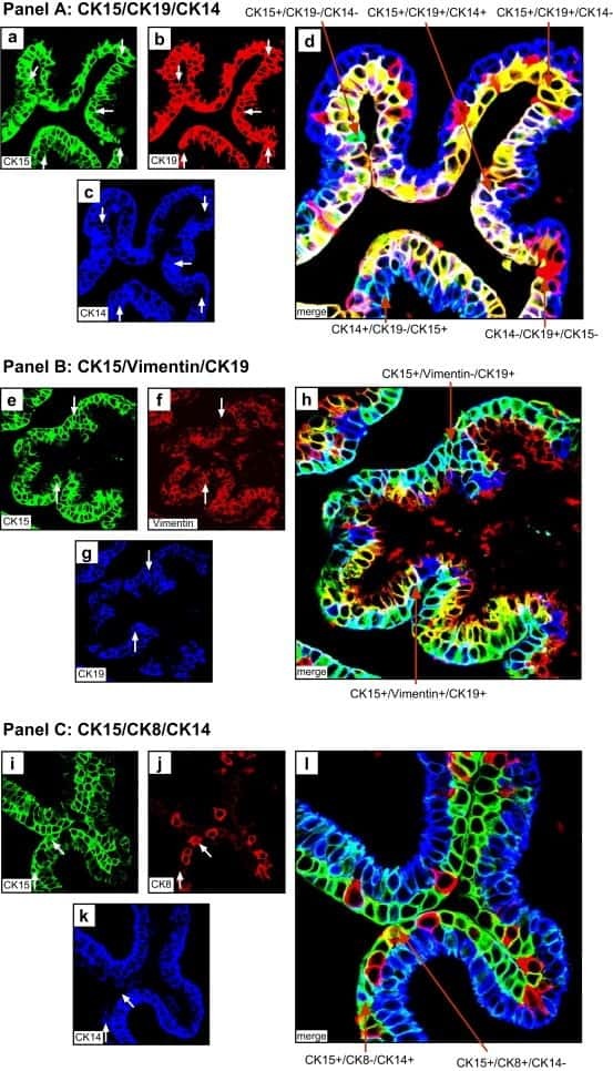
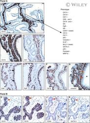
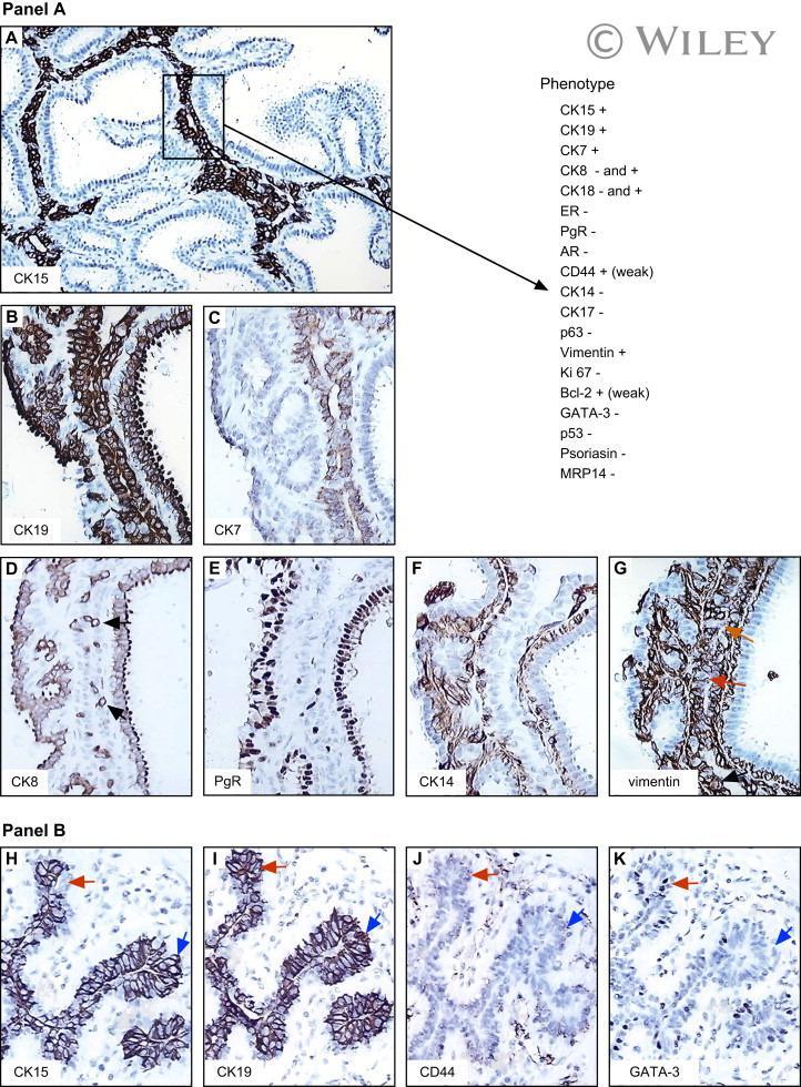
- Phenotype of non-malignant CK15+/CK19+ cells present in tumour 71. Panel A. Serial sections of paraffin-embedded tissue stained with antibodies against (A) CK15 (monoclonal), (B) CK19, (C) CK7, (D) CK8 (the black arrows indicate strongly positive cells), (E) (PgR), (F) CK14 and (G) vimentin. Only the area boxed in (A) is shown at larger magnification in figures B-G. The red arrows in G indicate vimentin negative cells. The phenotype of the cells is given at the top of Panel A. Panel B. Another area of the same section reacted with antibodies against CK15 (H), CK19 (I), CD44 (J), and GATA-3 (H). Blue arrows indicate CK15+/CK19+ cells, while the red arrows indicate CK15-/CK19+ cells which are differentiated as judged by the expression of GATA-3 (K).
- Submitted by
- Invitrogen Antibodies (provider)
- Main image

- Experimental details
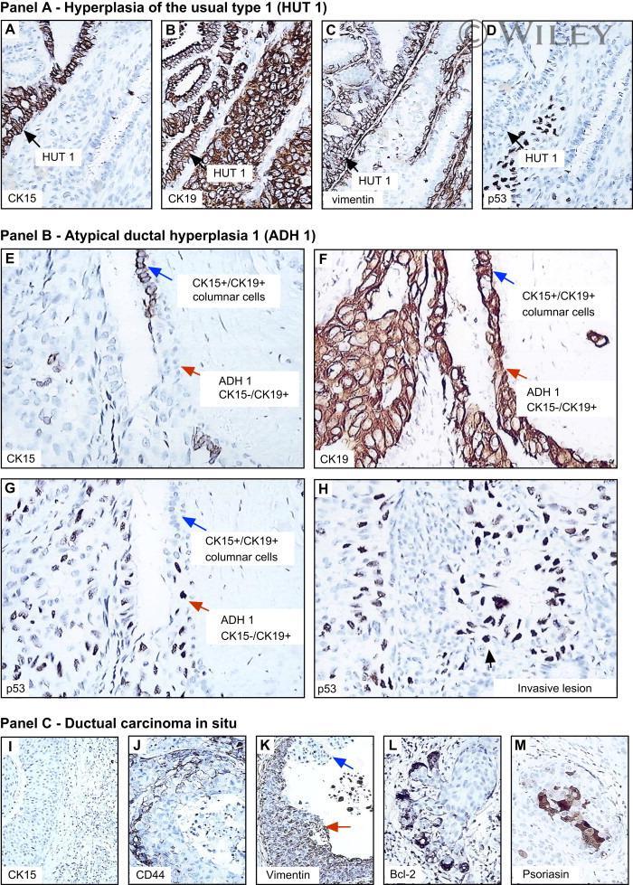
- IHC of non-malignant and premalignant lesions present in tumour 71. Panel A. Area with HUT 1 contiguous to CK15+/CK19+ cells stained with antibodies against CK15 (monoclonal) (A), CK19 (B), vimentin (C) and p53 (D). Panel B. ADH 1 contiguous to CK15+/CK19+ columnar cells reacted with antibodies against CK15 (E), CK19 (F), and p53 (G) (Table 2). For reference, (H) shows the invasive part of the tumour reacted with p53. Panel C. Ductal carcinoma in situ from T71 reacted with antibodies against CK15 (I), CD44 (J), vimentin (K), Bcl-2 (L), and psoriasin (S100 A7) (M). The red arrow in (K) indicates vimentin positive cells, while the blue one indicates negative ones.
- Submitted by
- Invitrogen Antibodies (provider)
- Main image
- Experimental details
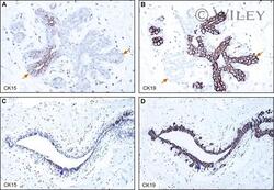
- CK15+ cells in normal TDLUs. Serial sections of normal ducts and lobules from biopsies of patients 14 (A, B) and 54 (C, D) stained with the CK15 (monoclonal) (A, C) and CK19 antibodies (B, D), respectively. Red arrows in (A) and (B) indicate cells that express either CK15 or CK19. Blue arrows in (C) and (D) indicate cells that co-express CK15 and CK19.
- Submitted by
- Invitrogen Antibodies (provider)
- Main image

- Experimental details
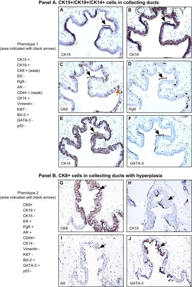
- CK15+/CK19+/CK14+ progenitor 1 cells in human large collecting ducts of the breast. Panel A. Serial sections of collecting ducts from patient 27 stained with antibodies against CK15 (polyclonal, Aviva) (A), CK19 (B), CK8 (C), PgR (D), CK14 (E) and GATA-3 (F). The phenotype of the progenitor cells (black arrows) in given for reference. The red arrow in (C) indicates a CK8 positive cell. Panel B. CK8 positive cells present in a large collecting duct of patient 27 with epithelial hyperplasia. Serial of collecting ducts were stained with antibodies against CK8 (G), CK15 (H), AR (I), and GATA-3 (J). The phenotype of the CK8 positive cells indicated with a black arrow is given for reference.
- Submitted by
- Invitrogen Antibodies (provider)
- Main image

- Experimental details
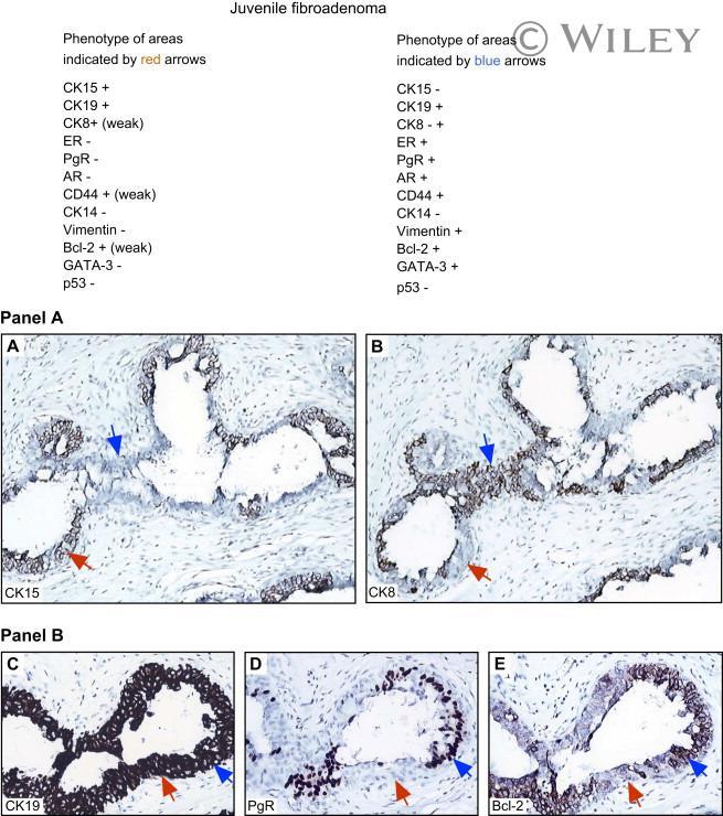
- CK15+/CK19+ cells present in the ducts of a juvenile fibroadenoma with epithelial hyperplasia. Panel A. Serial sections stained with the CK15 (polyclonal Aviva) (A) and CK8 antibodies (B) respectively. Red arrows indicate areas of the duct with CK15 positive cells, while blue ones indicated CK8 positive cells. Panel B. Serial sections of another area of the juvenile fibroadenoma stained with antibodies against CK19 (C), PgR (D), and Bcl-2 (E). Red arrows indicate areas of the duct with CK15+/CK8- cells, while blue ones indicate CK8+/CK15- cells. The extended phenotype of both cell types are given for reference.