 Explore
 Validate
 Learn
Learn Western blot
 ELISA
ELISAAntibody data
- Antibody Data
- Antigen structure
- References [0]
- Comments [0]
- Validations
- Western blot [4]
- Immunocytochemistry [1]
- Immunohistochemistry [1]
Submit
Validation data
Reference
Comment
Report error
- Product number
- MA5-15463 - Provider product page

- Provider
- Invitrogen Antibodies
- Product name
- Cytokeratin 19 Monoclonal Antibody (9H8G6)
- Antibody type
- Monoclonal
- Antigen
- Purifed from natural sources
- Description
- MA5-15463 targets Cytokeratin 19 in indirect ELISA, IHC and WB applications and shows reactivity with Human samples. The MA5-15463 immunogen is purified recombinant fragment of Cytokeratin 19 (aa80-400) expressed in E. Coli.. MA5-15463 detects Cytokeratin 19 which has a predicted molecular weight of approximately 44kDa.
- Reactivity
- Human
- Host
- Mouse
- Isotype
- IgG
- Antibody clone number
- 9H8G6
- Vial size
- 100 µL
- Concentration
- Conc. Not Determined
- Storage
- Store at 4°C short term. For long term storage, store at -20°C, avoiding freeze/thaw cycles.
No comments: Submit comment
Supportive validation
- Submitted by
- Invitrogen Antibodies (provider)
- Main image
- Experimental details
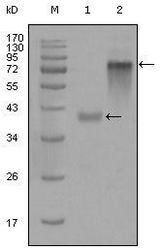
- Western blot analysis of Cytokeratin 19 using a Cytokeratin 19 monoclonal antibody (Product # MA5-15463) against a truncated KRT19-His recombinant protein (1) and full-length KRT19 (aa1-400) human IgG Fc transfected CHO-K1 cell lysate (2).

- Submitted by
- Invitrogen Antibodies (provider)
- Main image

- Experimental details
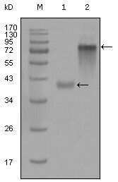
- Western blot analysis of Cytokeratin 19 using a Cytokeratin 19 monoclonal antibody (Product # MA5-15463) against a truncated KRT19-His recombinant protein (1) and full-length KRT19 (aa1-400) human IgG Fc transfected CHO-K1 cell lysate (2).
- Submitted by
- Invitrogen Antibodies (provider)
- Main image
- Experimental details
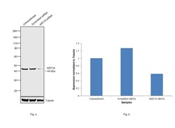
- Knockdown of KRT19 was achieved by transfecting MCF-7 with KRT19 specific siRNAs (Silencer® select Product # s7998). Western blot analysis (Fig. a) was performed using membrane enriched extracts from the KRT19 knockdown cells (lane 3), non-specific scrambled siRNA transfected cells (lane 2) and untransfected cells (lane 1). The blot was probed with Cytokeratin 19 Monoclonal Antibody (9H8G6) (Product # MA5-15463, 1:1000 dilution) and Goat anti-Mouse IgG (H+L), Superclonal™ Recombinant Secondary Antibody, HRP (Product # A28177, 0.25µg/ml, 1:4000 dilution). Densitometric analysis of this western blot is shown in histogram (Fig. b). Decrease in signal upon siRNA mediated knock down confirms that antibody is specific to KRT19.

- Submitted by
- Invitrogen Antibodies (provider)
- Main image
- Experimental details
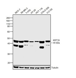
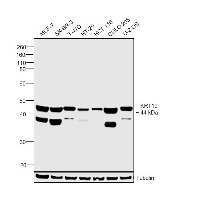
- Western blot was performed using Anti-Cytokeratin 19 Monoclonal Antibody (9H8G6) (Product # MA5-15463) and a 44 kDa band corresponding to KRT19 was observed across cell lines tested. An additional band around 38 kDa has been observed which is also reported for Cytokeratin 19. Membrane enriched extracts (30 µg lysate) of MCF-7 (Lane 1), SK-BR-3 (Lane 2), T-47D (Lane 3), HT-29 (Lane 4), HCT 116 (Lane 5), COLO 205 (Lane 6) and U-2 OS (Lane 7) were electrophoresed using NuPAGE™ 4-12% Bis-Tris Protein Gel (Product # NP0322BOX). Resolved proteins were then transferred onto a nitrocellulose membrane (Product # IB23001) by iBlot® 2 Dry Blotting System (Product # IB21001). The blot was probed with the primary antibody (1:1000 dilution) and detected by chemiluminescence with Goat anti-Mouse IgG (H+L) Superclonal™ Recombinant Secondary Antibody, HRP (Product # A28177, 1:4000 dilution) using the iBright FL 1000 (Product # A32752). Chemiluminescent detection was performed using Novex® ECL Chemiluminescent Substrate Reagent Kit (Product # WP20005).
Supportive validation
- Submitted by
- Invitrogen Antibodies (provider)
- Main image
- Experimental details
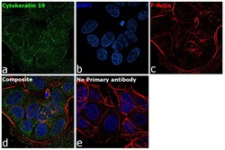
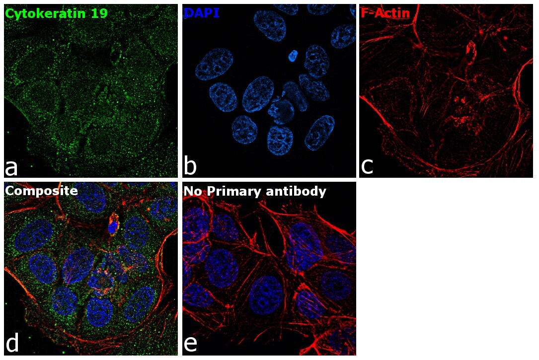
- Immunofluorescence analysis of Cytokeratin 19 was performed using 70% confluent log phase MCF-7 cells. The cells were fixed with 4% paraformaldehyde for 10 minutes, permeabilized with 0.1% Triton™ X-100 for 15 minutes, and blocked with 2% BSA for 1 hour at room temperature. The cells were labeled with Cytokeratin 19 Mouse Monoclonal Antibody (9H8G6) (Product # MA5-15463) at 1:100 dilution in 0.1% BSA, incubated at 4 degree Celsius overnight and then with Goat anti-Mouse IgG (H+L) Superclonal™ Recombinant Secondary Antibody, Alexa Fluor® 488 conjugate (Product # A28175) at a dilution of 1:2000 for 45 minutes at room temperature (Panel a: green). Nuclei (Panel b: blue) were stained with ProLong™ Diamond Antifade Mountant with DAPI (Product # P36962). F-actin (Panel c: red) was stained with Rhodamine Phalloidin (Product # R415, 1:300). Panel d represents the merged image showing cytoplasmic localization. Panel e represents control cells with no primary antibody to assess background. The images were captured at 60X magnification.
Supportive validation
- Submitted by
- Invitrogen Antibodies (provider)
- Main image
- Experimental details
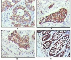
- Immunohistochemical analysis of paraffin-embedded human breast carcinoma (A), lung cancer (B) and normal colon tissue (C) using Cytokeratin 19 monoclonal antibody (Product # MA5-15463) followed with DAB staining
