 Explore
 Validate
 Learn
Learn Western blot
Antibody data
- Antibody Data
- Antigen structure
- References [8]
- Comments [0]
- Validations
- Western blot [2]
- Immunocytochemistry [2]
- Immunohistochemistry [1]
- Flow cytometry [1]
Submit
Validation data
Reference
Comment
Report error
- Product number
- MA5-12319 - Provider product page

- Provider
- Invitrogen Antibodies
- Product name
- Cytokeratin 19 Monoclonal Antibody (BA17)
- Antibody type
- Monoclonal
- Antigen
- Other
- Description
- MA5-12319 targets Cytokeratin 19 in IHC (P) and WB applications and shows reactivity with Human samples. The MA5-12319 immunogen is detergent soluble extract of human mammary epithelium.
- Reactivity
- Human
- Host
- Mouse
- Isotype
- IgG
- Antibody clone number
- BA17
- Vial size
- 500 µL
- Concentration
- 0.02 mg/mL
- Storage
- 4° C
Submitted references Histochemical and immunohistochemical characterization of chordoma in ferrets.
Anti-fibrotic effects of fresh and cryopreserved human amniotic membrane in a rat liver fibrosis model.
Ocular melanoma and mammary mucinous carcinoma in an African lion.
Characterizing and optimizing poly-L-lactide-co-ε-caprolactone membranes for urothelial tissue engineering.
Universal markers of thyroid malignancies: galectin-3, HBME-1, and cytokeratin-19.
Transplantation of allogeneic and xenogeneic placenta-derived cells reduces bleomycin-induced lung fibrosis.
An integrative computational model for intestinal tissue renewal.
Modulation by retinoic acid (RA) of squamous cell differentiation, cellular RA-binding proteins, and nuclear RA receptors in human head and neck squamous cell carcinoma cell lines.
Yui T, Ohmachi T, Matsuda K, Okamoto M, Taniyama H
The Journal of veterinary medical science 2015 Apr;77(4):467-73
The Journal of veterinary medical science 2015 Apr;77(4):467-73
Anti-fibrotic effects of fresh and cryopreserved human amniotic membrane in a rat liver fibrosis model.
Ricci E, Vanosi G, Lindenmair A, Hennerbichler S, Peterbauer-Scherb A, Wolbank S, Cargnoni A, Signoroni PB, Campagnol M, Gabriel C, Redl H, Parolini O
Cell and tissue banking 2013 Sep;14(3):475-88
Cell and tissue banking 2013 Sep;14(3):475-88
Ocular melanoma and mammary mucinous carcinoma in an African lion.
Cagnini DQ, Salgado BS, Linardi JL, Grandi F, Rocha RM, Rocha NS, Teixeira CR, Del Piero F, Sequeira JL
BMC veterinary research 2012 Sep 25;8:176
BMC veterinary research 2012 Sep 25;8:176
Characterizing and optimizing poly-L-lactide-co-ε-caprolactone membranes for urothelial tissue engineering.
Sartoneva R, Haaparanta AM, Lahdes-Vasama T, Mannerström B, Kellomäki M, Salomäki M, Sándor G, Seppänen R, Miettinen S, Haimi S
Journal of the Royal Society, Interface 2012 Dec 7;9(77):3444-54
Journal of the Royal Society, Interface 2012 Dec 7;9(77):3444-54
Universal markers of thyroid malignancies: galectin-3, HBME-1, and cytokeratin-19.
Barut F, Onak Kandemir N, Bektas S, Bahadir B, Keser S, Ozdamar SO
Endocrine pathology 2010 Jun;21(2):80-9
Endocrine pathology 2010 Jun;21(2):80-9
Transplantation of allogeneic and xenogeneic placenta-derived cells reduces bleomycin-induced lung fibrosis.
Cargnoni A, Gibelli L, Tosini A, Signoroni PB, Nassuato C, Arienti D, Lombardi G, Albertini A, Wengler GS, Parolini O
Cell transplantation 2009;18(4):405-22
Cell transplantation 2009;18(4):405-22
An integrative computational model for intestinal tissue renewal.
van Leeuwen IM, Mirams GR, Walter A, Fletcher A, Murray P, Osborne J, Varma S, Young SJ, Cooper J, Doyle B, Pitt-Francis J, Momtahan L, Pathmanathan P, Whiteley JP, Chapman SJ, Gavaghan DJ, Jensen OE, King JR, Maini PK, Waters SL, Byrne HM
Cell proliferation 2009 Oct;42(5):617-36
Cell proliferation 2009 Oct;42(5):617-36
Modulation by retinoic acid (RA) of squamous cell differentiation, cellular RA-binding proteins, and nuclear RA receptors in human head and neck squamous cell carcinoma cell lines.
Zou CP, Clifford JL, Xu XC, Sacks PG, Chambon P, Hong WK, Lotan R
Cancer research 1994 Oct 15;54(20):5479-87
Cancer research 1994 Oct 15;54(20):5479-87
No comments: Submit comment
Supportive validation
- Submitted by
- Invitrogen Antibodies (provider)
- Main image
- Experimental details
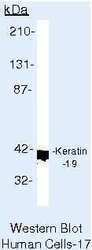
- Western blot of Cytokeratin 19 using Cytokeratin 19 Monoclonal Antibody (Product # MA5-12319) on LS174T Cells.

- Submitted by
- Invitrogen Antibodies (provider)
- Main image
- Experimental details
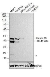
- Western blot was performed using Anti-Cytokeratin 19 Monoclonal Antibody (BA17) (Product # MA5-12319) and 38-44kDa bands corresponding to Cytokeratin 19 was observed across cell lines tested except for SH-SY5Y and HEK-293 cells which are reported to be low to negative for expression of Cytokeratin 19. An additional faint non-specific band (*) was observed in MCF7. Whole cell extracts (15 µg lysate) of MCF7 (Lane 1), SK-BR-3 (Lane 2), MDA-MB-231 (Lane 3), SH-SY5Y (Lane 4) and HEK-293 (Lane 5) were electrophoresed using NuPAGE™ 4-12% Bis-Tris Protein Gel (Product # NP0322BOX). Resolved proteins were then transferred onto a Nitrocellulose membrane (Product # IB23002) by iBlot® 2 Dry Blotting System (Product # IB21001). The blot was probed with the primary antibody (1ug/ml) and detected by chemiluminescence with Goat anti-Mouse IgG (H+L) Superclonal™ Recombinant Secondary Antibody, HRP (Product # A28177,1:4000 dilution) using the iBright FL 1000 (Product # A32752). Chemiluminescent detection was performed using Novex® ECL Chemiluminescent Substrate Reagent Kit (Product # WP20005).

Supportive validation
- Submitted by
- Invitrogen Antibodies (provider)
- Main image
- Experimental details
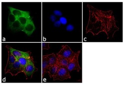
- Immunofluorescent analysis of Cytokeratin 19 was performed using 70% confluent log phase MCF-7 cells. The cells were fixed with 4% paraformaldehyde for 10 minutes, permeabilized with 0.1% Triton™ X-100 for 10 minutes, and blocked with 1% BSA for 1 hour at room temperature. The cells were labeled with Cytokeratin 19 (BA17) Mouse Monoclonal Antibody (Product # MA5-12319) at 2 µg/mL in 0.1% BSA and incubated for 3 hours at room temperature and then labeled with Goat anti-Mouse IgG (H+L) Superclonal™ Secondary Antibody, Alexa Fluor® 488 conjugate (Product # A28175) a dilution of 1:2000 for 45 minutes at room temperature (Panel a: green). Nuclei (Panel b: blue) were stained with SlowFade® Gold Antifade Mountant with DAPI (Product # S36938). F-actin (Panel c: red) was stained with Alexa Fluor® 555 Rhodamine Phalloidin (Product # R415, 1:300). Panel d represents the merged image showing cytoplasmic localization. Panel e shows the no primary antibody control. The images were captured at 60X magnification.

- Submitted by
- Invitrogen Antibodies (provider)
- Main image
- Experimental details
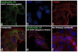
- Immunofluorescence analysis of Cytokeratin 19 was performed using 70% confluent log phase MCF7 and SH-SY5Y cells. The cells were fixed with 4% paraformaldehyde for 10 minutes, permeabilized with 0.1% Triton™ X-100 for 15 minutes, and blocked with 2% BSA for 45 minutes at room temperature. The cells were labeled with Cytokeratin 19 Monoclonal Antibody (BA17) (Product # MA5-12319) at 2 µg/mL in 0.1% BSA, incubated at 4 degree celsius overnight and then labeled with Donkey anti-Mouse IgG (H+L) Highly Cross-Adsorbed Secondary Antibody, Alexa Fluor Plus 488 (Product # A32766), (1:2000 dilution), for 45 minutes at room temperature (Panel a: Green). Nuclei (Panel b: Blue) were stained with ProLong™ Diamond Antifade Mountant with DAPI (Product # P36962). F-actin (Panel c: Red) was stained with Rhodamine Phalloidin (Product # R415, 1:300). Panel d represents the merged image showing cytoskeletal localization in MCF7 cells but not in SH-SY5Y cells (Panel e) which is reported to be low to negative for Cytokeratin 19 expression. Panel f represents control cells with no primary antibody to assess the background. The images were captured at 60X magnification.

Supportive validation
- Submitted by
- Invitrogen Antibodies (provider)
- Main image

- Experimental details
- Formalin-fixed, paraffin-embedded human colon carcinoma stained with Keratin-19 antibody using peroxidase-conjugate and AEC chromogen. Note cytoplasmic staining of tumor cells.

Supportive validation
- Submitted by
- Invitrogen Antibodies (provider)
- Main image

- Experimental details
- Flow cytometry analysis of Cytokeratin 19 was done on MCF7 cells. Cells were fixed with 70% ethanol for 10 minutes, permeabilized with 0.25% Triton™ X-100 for 20 minutes, and blocked with 5% BSA for 30 minutes at room temperature. Cells were labeled with Cytokeratin 19 Mouse Monoclonal Antibody (MA5-12319, red histogram) or with mouse isotype control (pink histogram) at 3-5 ug/million cells in 2.5% BSA. After incubation at room temperature for 2 hours, the cells were labeled with Alexa Fluor® 488 Rabbit Anti-Mouse Secondary Antibody (A11059) at a dilution of 1:400 for 30 minutes at room temperature. The representative 10, 000 cells were acquired and analyzed for each sample using an Attune® Acoustic Focusing Cytometer. The purple histogram represents unstained control cells and the green histogram represents no-primary-antibody control.
