 Explore
 Validate
 Learn
Learn Western blot
Antibody data
- Antibody Data
- Antigen structure
- References [1]
- Comments [0]
- Validations
- Western blot [3]
- Immunocytochemistry [2]
- Immunohistochemistry [3]
- Other assay [2]
Submit
Validation data
Reference
Comment
Report error
- Product number
- PA5-29582 - Provider product page

- Provider
- Invitrogen Antibodies
- Product name
- Cytokeratin 19 Polyclonal Antibody
- Antibody type
- Polyclonal
- Antigen
- Recombinant protein fragment
- Description
- Recommended positive controls: HeLa, HepG2. Predicted reactivity: Mouse (91%), Rat (93%), Dog (92%), Chicken (84%), Bovine (92%). Store product as a concentrated solution. Centrifuge briefly prior to opening the vial.
- Reactivity
- Human
- Host
- Rabbit
- Isotype
- IgG
- Vial size
- 100 µL
- Concentration
- 1 mg/mL
- Storage
- Store at 4°C short term. For long term storage, store at -20°C, avoiding freeze/thaw cycles.
Submitted references Concurrent isolation of hepatic stem cells and hepatocytes from the human liver.
Lee SML, Bertinetti-Lapatki C, Schiergens TS, Jauch KW, Roth AB, Thasler WE
In vitro cellular & developmental biology. Animal 2020 Mar;56(3):253-260
In vitro cellular & developmental biology. Animal 2020 Mar;56(3):253-260
No comments: Submit comment
Supportive validation
- Submitted by
- Invitrogen Antibodies (provider)
- Main image
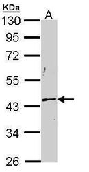
- Experimental details
- Western blot analysis of Cytokeratin 19 using 30 µg of HepG2 lysate. Samples were loaded onto a 10% SDS-PAGE gel and probed with a Cytokeratin 19 polyclonal antibody (Product # PA5-29582) at a dilution of 1:1000.

- Submitted by
- Invitrogen Antibodies (provider)
- Main image
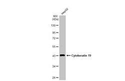
- Experimental details
- Western Blot using Cytokeratin 19 Polyclonal Antibody (Product # PA5-29582). Whole cell extract (30 µg) was separated by 10% SDS-PAGE, and the membrane was blotted with Cytokeratin 19 Polyclonal Antibody (Product # PA5-29582) diluted at 1:1,000. The HRP-conjugated anti-rabbit IgG antibody was used to detect the primary antibody.

- Submitted by
- Invitrogen Antibodies (provider)
- Main image
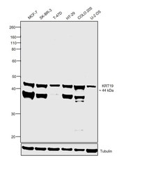
- Experimental details
- Western blot was performed using Anti-Cytokeratin 19 Polyclonal Antibody (Product # PA5-29582) and a 44 kDa band corresponding to KRT19 was observed across cell lines tested. An additional band around 38 kDa has been observed which is reported for Cytokeratin 19 expression. Membrane enriched extracts (30 µg lysate) of MCF-7 (Lane 1), SK-BR-3 (Lane 2), T-47D (Lane 3), HT-29 (Lane 4), COLO 205 (Lane 5) and U-2 OS (Lane 6) were electrophoresed using NuPAGE™ 4-12% Bis-Tris Protein Gel (Product # NP0322BOX). Resolved proteins were then transferred onto a nitrocellulose membrane (Product # IB23001) by iBlot® 2 Dry Blotting System (Product # IB21001). The blot was probed with the primary antibody (1:2000 dilution) and detected by chemiluminescence with Goat anti-Rabbit IgG (H+L) Superclonal™ Recombinant Secondary Antibody, HRP (Product # A27036, 1:4000 dilution) using the iBright FL 1000 (Product # A32752). Chemiluminescent detection was performed using Novex® ECL Chemiluminescent Substrate Reagent Kit (Product # WP20005).

Supportive validation
- Submitted by
- Invitrogen Antibodies (provider)
- Main image
- Experimental details
- Immunofluorescent analysis of Cytokeratin 19 in paraformaldehyde-fixed HeLa cells using a Cytokeratin 19 polyclonal antibody (Product # PA5-29582) at a 1:200 dilution.

- Submitted by
- Invitrogen Antibodies (provider)
- Main image
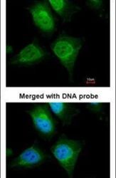
- Experimental details
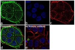
- Immunofluorescence analysis of Cytokeratin 19 was performed using 70% confluent log phase MCF7 cells. The cells were fixed with 4% paraformaldehyde for 10 minutes, permeabilized with 0.1% Triton™ X-100 for 15 minutes, and blocked with 2% BSA for 1 hour at room temperature. The cells were labeled with Cytokeratin 19 Rabbit Polyclonal Antibody (Product # PA5-29582) at 5 µg/mL in 0.1% BSA, incubated at 4 degree Celsius overnight and then labeled with Goat anti-Rabbit IgG (H+L), Superclonal™ Recombinant Secondary Antibody, Alexa Fluor 488 (Product # A27034) at a dilution of 1:2000 for 45 minutes at room temperature (Panel a: green). Nuclei (Panel b: blue) were stained with ProLong™ Diamond Antifade Mountant with DAPI (Product # P36962). F-actin (Panel c: red) was stained with Rhodamine Phalloidin (Product # R415). Panel d represents the merged image showing cytoplasmic localization. Panel e represents control cells with no primary antibody to assess background. The images were captured at 60X magnification.

Supportive validation
- Submitted by
- Invitrogen Antibodies (provider)
- Main image

- Experimental details
- Immunohistochemistry (Paraffin) analysis of Cytokeratin 19 was performed in paraffin-embedded human lung adenocarcinoma tissue using Cytokeratin 19 Polyclonal Antibody (Product # PA5-29582) at a dilution of 1:500.

- Submitted by
- Invitrogen Antibodies (provider)
- Main image

- Experimental details
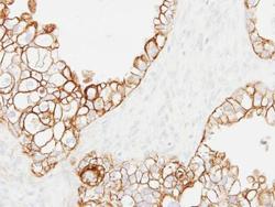
- Cytokeratin 19 Polyclonal Antibody detects Cytokeratin 19 protein at cell membrane by immunohistochemical analysis. Sample: Paraffin-embedded mouse tongue. Cytokeratin 19 stained by Cytokeratin 19 Polyclonal Antibody (Product # PA5-29582) diluted at 1:500. Antigen Retrieval: Citrate buffer, pH 6.0, 15 min.

- Submitted by
- Invitrogen Antibodies (provider)
- Main image
- Experimental details
- Immunohistochemical analysis of paraffin-embedded OVCA xenograft, using Cytokeratin 19 (Product # PA5-29582) antibody at 1:500 dilution. Antigen Retrieval: Citrate buffer, pH 6.0, 15 min.

Supportive validation
- Submitted by
- Invitrogen Antibodies (provider)
- Main image

- Experimental details
- Figure 1. EpCAM and CK19 colocalise in human liver. Immunofluorescent staining of EpCAM and CK19 in cirrhotic ( a - c ) or in macroscopically normal liver ( d - f ). EpCAM localisation is visualised in red in panels ( a ) and ( d ), while CK19 localisation is visualised in green in panels ( b ) and ( e ). Co-localised expression of EpCAM and CK19 can be seen in panel ( c ) for cirrhotic liver and panel ( f ) for normal liver. Scale bars in the figure indicate a length 100 mum.

- Submitted by
- Invitrogen Antibodies (provider)
- Main image

- Experimental details
- Figure 4. Hepatic stem cell characterisation. Immunofluorescent labelling of ( a ) albumin (green) and ( b ) CK19 (red) in hepatic stem cells 5 d after isolation with the co-localised expression of albumin and CK19 shown in panel ( c ). The corresponding secondary antibody control image is shown in panel ( d ). These cells express ( e ) proliferation marker Ki67 (green) in the nuclei making them appear cyan when compared with the corresponding ( f ) secondary antibody control with blue nuclei. Nuclei are counterstained blue with DAPI. Scale bars in the figure indicate a length 100 mum.
