 Explore
 Validate
 Learn
Learn Western blot
Antibody data
- Antibody Data
- Antigen structure
- References [0]
- Comments [0]
- Validations
- Western blot [1]
- Immunocytochemistry [1]
- Immunoprecipitation [1]
- Immunohistochemistry [1]
Submit
Validation data
Reference
Comment
Report error
- Product number
- TA328744 - Provider product page

- Provider
- OriGene
- Product name
- Rabbit Polyclonal Anti-TRPA1 (extracellular)
- Antibody type
- Polyclonal
- Description
- Rabbit Polyclonal Anti-TRPA1 (extracellular)
- Host
- Rabbit
- Conjugate
- Unconjugated
- Epitope
- TRPA1
- Antibody clone number
- NULL
- Vial size
- 200 µl
- Concentration
- NULL
No comments: Submit comment
Supportive validation
- Submitted by
- OriGene (provider)
- Main image
- Experimental details
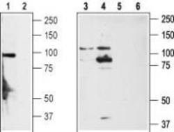
- Western blot analysis of rat DRG (lanes 1,2), non-differentiated PC12 cells (lanes 3,5) and differentiated PC12 cells (lanes 4,6) lysates: 1,3,4. Anti-TRPA1 (extracellular) antibody, (1:200). 2,5,6. Anti-TRPA1 (extracellular) antibody, preincubated with the control peptide antigen.
- Validation comment
- WB

Supportive validation
- Submitted by
- OriGene (provider)
- Main image

- Experimental details
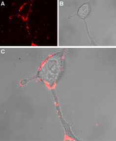
- Expression of TRPA1 in rat PC12 cells. Immunocytochemical staining of intact living rat PC12 cells. A. Extracellular staining of cells using Anti-TRPA1 (extracellular) antibody, (1:50) followed by goat anti-rabbit-AlexaFluor-594 secondary antibody (red). B. Live view of the cells. C. Merged image of A and B.
- Validation comment
- IF
Supportive validation
- Submitted by
- OriGene (provider)
- Main image

- Experimental details
- Immunoprecipitation of PC-12 lysates: 1. PC-12 lysates. 2. PC-12 lysates + Anti-TRPA1 (extracellular) antibody (6.5 ug) + protein A beads. 3. Anti-TRPA1 (extracellular) antibody + protein A beads.
- Validation comment
- IP

Supportive validation
- Submitted by
- OriGene (provider)
- Main image
- Experimental details
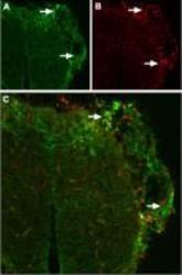
- Expression of TRPA1 in mouse DRG. Immunohistochemical staining of mouse dorsal root ganglion (DRG) frozen sections using Anti-TRPA1 (extracellular) antibody. A. TRPA1 (green) was distributed in patches (arrows). B. Neurons containing neurofilament 200 (red) also were distributed in patches. C. Confocal merge of TRPA1 and neurofilament 200 demonstrate partial overlap of these patches (arrows).
- Validation comment
- IHC
