Explore
 Validate
 Learn
Learn Western blot
Antibody data
- Antibody Data
- Antigen structure
- References [0]
- Comments [0]
- Validations
- Western blot [3]
- Immunocytochemistry [1]
- Immunohistochemistry [1]
Submit
Validation data
Reference
Comment
Report error
- Product number
- MA1-802 - Provider product page

- Provider
- Invitrogen Antibodies
- Product name
- TPX2 Monoclonal Antibody (18D5-1)
- Antibody type
- Monoclonal
- Antigen
- Recombinant full-length protein
- Description
- MA1-802 detects the TPX2 in human, rat and mouse cells.
- Antibody clone number
- 18D5-1
- Concentration
- 1 mg/mL
No comments: Submit comment
Supportive validation
- Submitted by
- Invitrogen Antibodies (provider)
- Main image
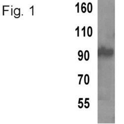
- Experimental details
- Western blot analysis of TPX2 using a monoclonal antibody (Product # MA1-802).
- Submitted by
- Invitrogen Antibodies (provider)
- Main image
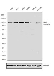
- Experimental details
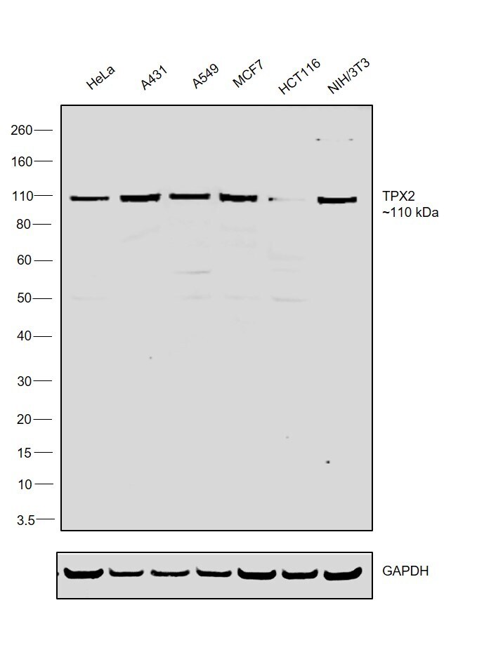
- Western blot was performed using Anti-TPX2 Monoclonal Antibody (18D5-1) (Product # MA1-802) and a ~110 kDa band corresponding to Targeting protein for Xklp2 was observed across cell lines tested. Nuclear enriched extracts (30 µg lysate) of HeLa (Lane 1), A431 (Lane 2), A549 (Lane 3), MCF7 (Lane 4), HCT116 (Lane 5) and NIH/3T3 ((Lane 6) were electrophoresed using NuPAGE™ 4-12% Bis-Tris Protein Gel (Product # NP0321BOX). Resolved proteins were then transferred onto a nitrocellulose membrane (Product # IB23001) by iBlot® 2 Dry Blotting System (Product # IB21001). The blot was probed with the primary antibody (1 µg/mL) and detected by chemiluminescence with Goat anti-Mouse IgG (H+L) Superclonal™ Recombinant Secondary Antibody, HRP (Product # A28177,1:20000 dilution) using the iBright™ FL1500 Imaging System (Product # A44115). Chemiluminescent detection was performed using SuperSignal™ West Dura Extended Duration Substrate (Product # 34076).
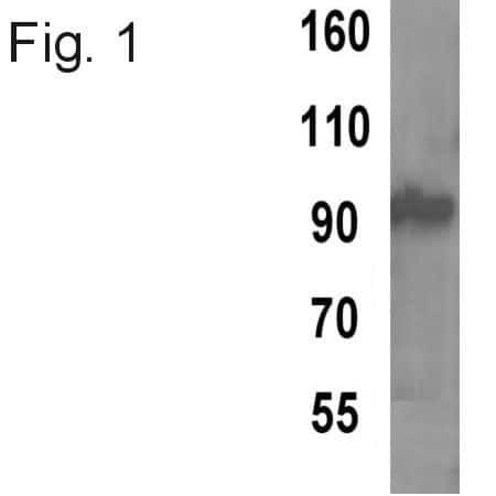
- Submitted by
- Invitrogen Antibodies (provider)
- Main image
- Experimental details
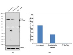
- Knockdown of Targeting protein for Xklp2 was achieved by transfecting HeLa with Targeting protein for Xklp2 specific siRNAs (Silencer® select Product # S22745, S22746). Western blot analysis (Fig. a) was performed using Nuclear enriched extracts from the Targeting protein for Xklp2 knockdown cells (lane 3), non-targeting scrambled siRNA transfected cells (lane 2) and untransfected cells (lane 1). The blot was probed with TPX2 Monoclonal Antibody (18D5-1) (Product # MA1-802, 1 µg/mL ) and Goat anti-Mouse IgG (H+L) Superclonal™ Recombinant Secondary Antibody, HRP (Product # A28177, 1:20000 dilution). Densitometric analysis of this western blot is shown in histogram (Fig. b). Decrease in signal upon siRNA mediated knock down confirms that antibody is specific to Targeting protein for Xklp2.

Supportive validation
- Submitted by
- Invitrogen Antibodies (provider)
- Main image

- Experimental details
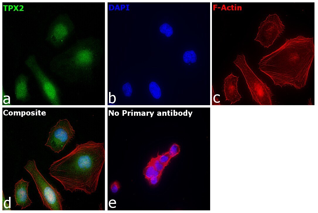
- Immunofluorescence analysis of Targeting protein for Xklp2 was performed using 70% confluent log phase HeLa cells. The cells were fixed with 4% paraformaldehyde for 10 minutes, permeabilized with 0.1% Triton™ X-100 for 10 minutes, and blocked with 2% BSA for 1 hour at room temperature. The cells were labeled with TPX2 Monoclonal Antibody (18D5-1) (Product # MA1-802) at 1:200 dilution in 0.1% BSA, incubated at 4 degree celsius overnight and then labeled with Goat anti-Mouse IgG (H+L) Highly Cross-Adsorbed Secondary Antibody, Alexa Fluor Plus 488 (Product # A32723), (1:2000 dilution), for 45 minutes at room temperature (Panel a: Green). Nuclei (Panel b:Blue) were stained with ProLong™ Diamond Antifade Mountant with DAPI (Product # P36962). F-actin (Panel c: Red) was stained with Rhodamine Phalloidin (Product # R415, 1:300). Panel d represents the merged image showing Nuclear localization. Panel e represents control cells with no primary antibody to assess background. The images were captured at 60X magnification in EVOS™ M7000 Imaging System (Product # AMF7000) and externally deconvoluted (D.Sage et al. / Methods 115 (2017) 28–41).
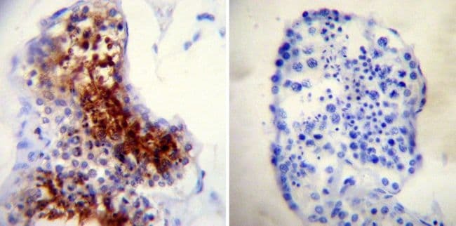
Supportive validation
- Submitted by
- Invitrogen Antibodies (provider)
- Main image

- Experimental details
- Immunohistochemistry was performed on biopsies of deparaffinized Human testis tissue. To expose target proteins, heat induced antigen retrieval was performed using 10mM sodium citrate (pH6.0) buffer, microwaved for 8-15 minutes. Following antigen retrieval tissues were blocked in 3% BSA-PBS for 30 minutes at room temperature. Tissues were then probed at a dilution of 1:200 with a mouse monoclonal antibody recognizing TPX2 (Product # MA1-802) or without primary antibody (negative control) overnight at 4°C in a humidified chamber. Tissues were washed extensively with PBST and endogenous peroxidase activity was quenched with a peroxidase suppressor. Detection was performed using a biotin-conjugated secondary antibody and SA-HRP, followed by colorimetric detection using DAB. Tissues were counterstained with hematoxylin and prepped for mounting.