Explore
 Validate
 Learn
Learn Western blot
 Immunocytochemistry
 Immunoprecipitation
Antibody data
- Antibody Data
- Antigen structure
- References [1]
- Comments [0]
- Validations
- Immunocytochemistry [2]
- Other assay [1]
Submit
Validation data
Reference
Comment
Report error
- Product number
- PA3-16832 - Provider product page

- Provider
- Invitrogen Antibodies
- Product name
- TPX2 Polyclonal Antibody
- Antibody type
- Polyclonal
- Antigen
- Recombinant full-length protein
- Description
- Suggested positive control: antigen standard for TPX2 (transient overexpression lysate).
- Reactivity
- Human, Mouse, Rat, Bovine, Porcine
- Host
- Rabbit
- Isotype
- IgG
- Vial size
- 100 μL
- Concentration
- Conc. Not Determined
- Storage
- -20°C, Avoid Freeze/Thaw Cycles
Submitted references HSP70 regulates Eg5 distribution within the mitotic spindle and modulates the cytotoxicity of Eg5 inhibitors.
Fang CT, Kuo HH, Hsu SC, Yih LH
Cell death & disease 2020 Sep 1;11(8):715
Cell death & disease 2020 Sep 1;11(8):715
No comments: Submit comment
Supportive validation
- Submitted by
- Invitrogen Antibodies (provider)
- Main image

- Experimental details
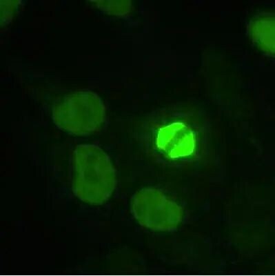
- Immunocytochemistry analysis of TPX2 in HeLa cells fixed in 3.5% paraformaldehyde. Samples were incubated in TPX2 polyclonal antibody (Product # PA3-16832) using a dilution of 1:1000. Nuclear staining during interphase and spindle staining during mitosis.

- Submitted by
- Invitrogen Antibodies (provider)
- Main image

- Experimental details
- Immunocytochemistry analysis of TPX2 in HeLa cells fixed in 3.5% paraformaldehyde. Samples were incubated in TPX2 polyclonal antibody (Product # PA3-16832) using a dilution of 1:1000. Nuclear staining during interphase and spindle staining during mitosis.
Supportive validation
- Submitted by
- Invitrogen Antibodies (provider)
- Main image
- Experimental details
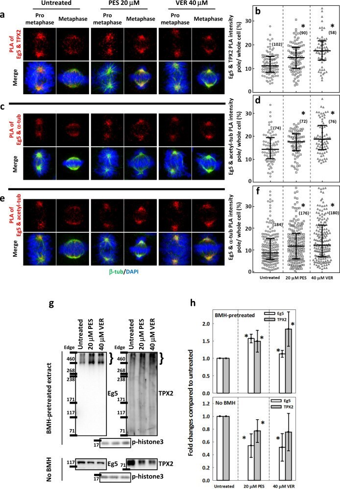
- Fig. 5 HSP70 is required for proper Eg5 interactions with TPX2, acetylated tubulin and alpha-tubulin. Representative images of Eg5-TPX2 PLA ( a ), Eg5-alpha-tubulin PLA ( c ) and Eg5-acetyl-tubulin PLA ( e ) signals are shown in red in untreated and PES- or VER-treated prometaphase and metaphase cells counterstained with beta-tubulin (green) and DAPI (blue). The ratio of the PLA signal at the poles to the whole-cell signal was measured as described, and the interquartile distributions from two independent experiments are shown for ( b ) Eg5-TPX2, ( d ) Eg5-alpha-tubulin, and ( f ) Eg5-acetyl-tubulin. The numbers in parentheses indicate the number of the cells measured. * p < 0.05 compared to untreated by Mann-Whitney Rank Sum test. g (upper) BMH-mediated crosslinking was performed as described, revealing a protein complex >460 kD containing Eg5 and TPX2 in synchronized mitotic cells that were untreated or treated with PES or VER. (Lower) Total Eg5 and TPX2 proteins in the denatured lysate. h The bar charts show the levels of Eg5 and TPX2 compared to the untreated cells, as normalized by the mitosis marker p-histone3 for BMH-crosslinked samples (upper) and no BMH-treated samples (lower). Mean +- SD of six replicates from three independent experiments is shown. * p < 0.05 compared to untreated by Student's t test.