Explore
 Validate
 Learn
Learn Western blot
 Immunocytochemistry
Antibody data
- Antibody Data
- Antigen structure
- References [1]
- Comments [0]
- Validations
- Immunocytochemistry [2]
- Immunoprecipitation [1]
- Other assay [4]
Submit
Validation data
Reference
Comment
Report error
- Product number
- PA5-80842 - Provider product page

- Provider
- Invitrogen Antibodies
- Product name
- NCAPH Polyclonal Antibody
- Antibody type
- Polyclonal
- Antigen
- Synthetic peptide
- Description
- This product is preservative free. It is recommended to add sodium azide to avoid contamination (final concentration 0.05%-0.1%). This antibody has specificity for Human NCAPH.
- Reactivity
- Human
- Host
- Rabbit
- Isotype
- IgG
- Vial size
- 100 μL
- Concentration
- 1 mg/mL
- Storage
- Store at 4°C short term. For long term storage, store at -20°C, avoiding freeze/thaw cycles.
Submitted references Non-SMC condensin I complex subunit H promotes the malignant progression and cisplatin resistance of breast cancer MCF-7 cells.
Liao L, Cheng H, Liu S
Oncology letters 2022 Sep;24(3):317
Oncology letters 2022 Sep;24(3):317
No comments: Submit comment
Supportive validation
- Submitted by
- Invitrogen Antibodies (provider)
- Main image

- Experimental details
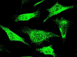
- Immunofluorescence staining of NCAPH in HeLa cells. Cells were fixed with 4% PFA, permeabilzed with 0.3% Triton X-100 in PBS, blocked with 10% serum, and incubated with NCAPH Polyclonal Antibody (Product # PA5-80842, 1:1,000) at 4°C overnight. Then cells were stained with the Alexa Fluor®488-conjugated Goat Anti-rabbit IgG secondary antibody (green). Positive staining was localized to cytoplasm and nucleus.

- Submitted by
- Invitrogen Antibodies (provider)
- Main image
- Experimental details
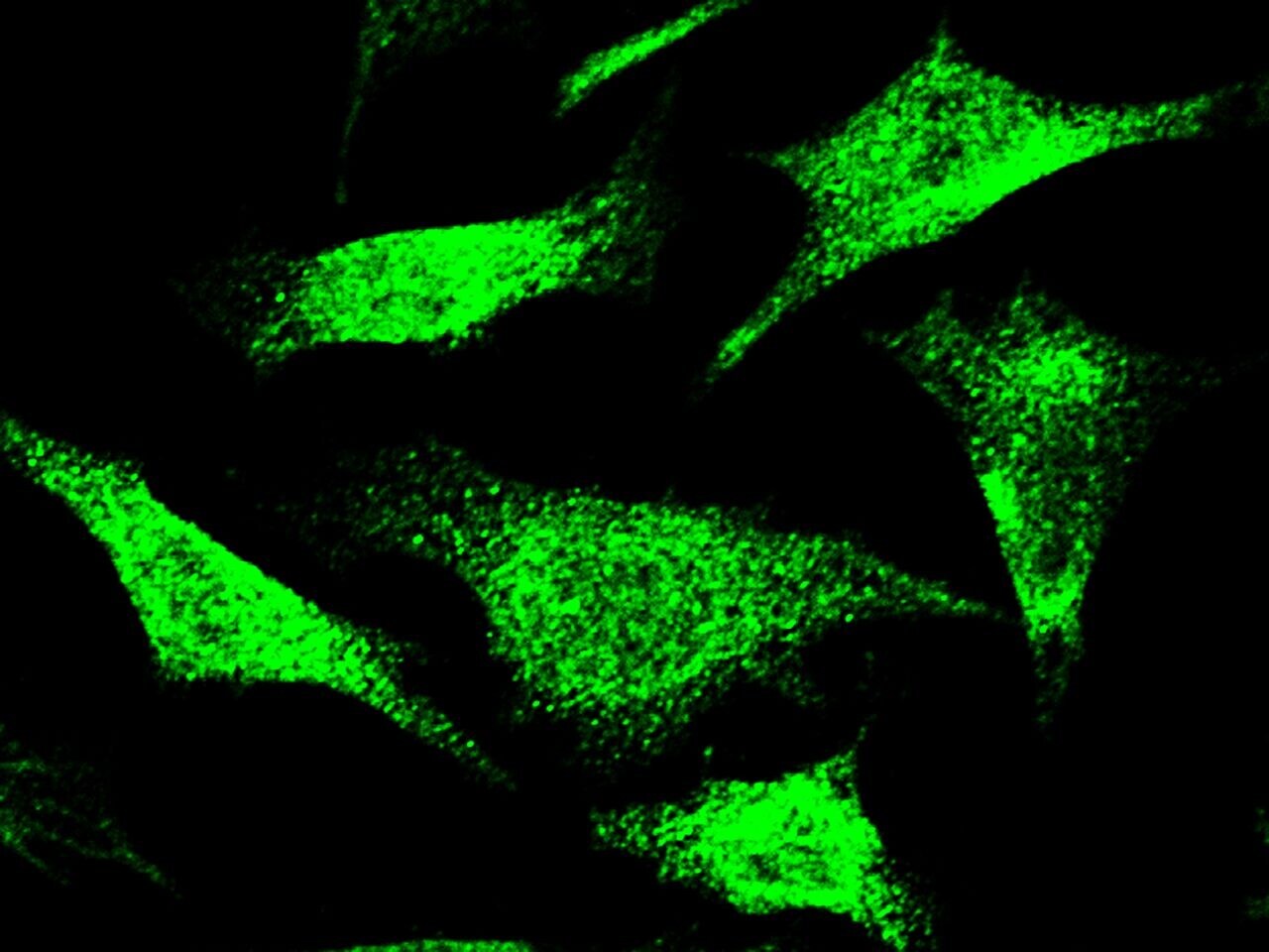
- Immunofluorescence staining of NCAPH in HeLa cells. Cells were fixed with 4% PFA, permeabilzed with 0.3% Triton X-100 in PBS, blocked with 10% serum, and incubated with NCAPH Polyclonal Antibody (Product # PA5-80842, 1:1,000) at 4°C overnight. Then cells were stained with the Alexa Fluor®488-conjugated Goat Anti-rabbit IgG secondary antibody (green). Positive staining was localized to cytoplasm and nucleus.
Supportive validation
- Submitted by
- Invitrogen Antibodies (provider)
- Main image
- Experimental details
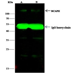
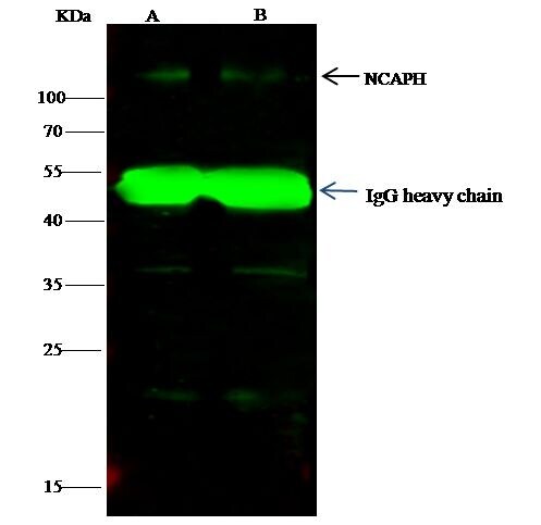
- NCAPH Immunoprecipitation using: Lane A: 0.5 mg K562 Whole Cell Lysate, Lane B: 0.5 mg HepG2 Whole Cell Lysate 4 µL with NCAPH Polyclonal Antibody (Product # PA5-80842) and 15 µL of 50 % Protein G agarose. Primary antibody: NCAPH Polyclonal Antibody, at 1:100 dilution. Secondary antibody: Dylight 800-labeled antibody to rabbit IgG (H+L), at 1:5,000 dilution. Developed using the Odyssey technique. Performed under reducing conditions. Predicted band size: 83 kDa. Observed band size: 110 kDa.

Supportive validation
- Submitted by
- Invitrogen Antibodies (provider)
- Main image

- Experimental details
- NCAPH Immunoprecipitation using: Lane A: 0.5 mg K562 Whole Cell Lysate, Lane B: 0.5 mg HepG2 Whole Cell Lysate 4 µL with NCAPH Polyclonal Antibody (Product # PA5-80842) and 15 µL of 50 % Protein G agarose. Primary antibody: NCAPH Polyclonal Antibody, at 1:100 dilution. Secondary antibody: Dylight 800-labeled antibody to rabbit IgG (H+L), at 1:5,000 dilution. Developed using the Odyssey technique. Performed under reducing conditions. Predicted band size: 83 kDa. Observed band size: 110 kDa.
- Submitted by
- Invitrogen Antibodies (provider)
- Main image
- Experimental details
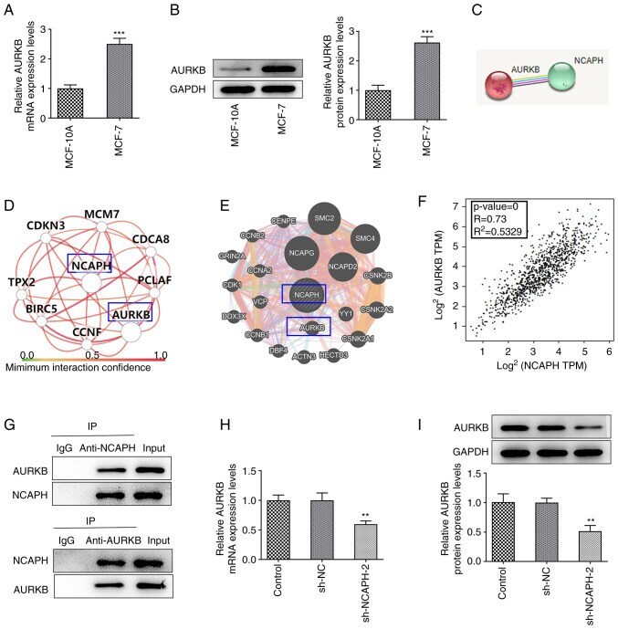
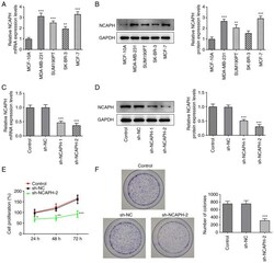
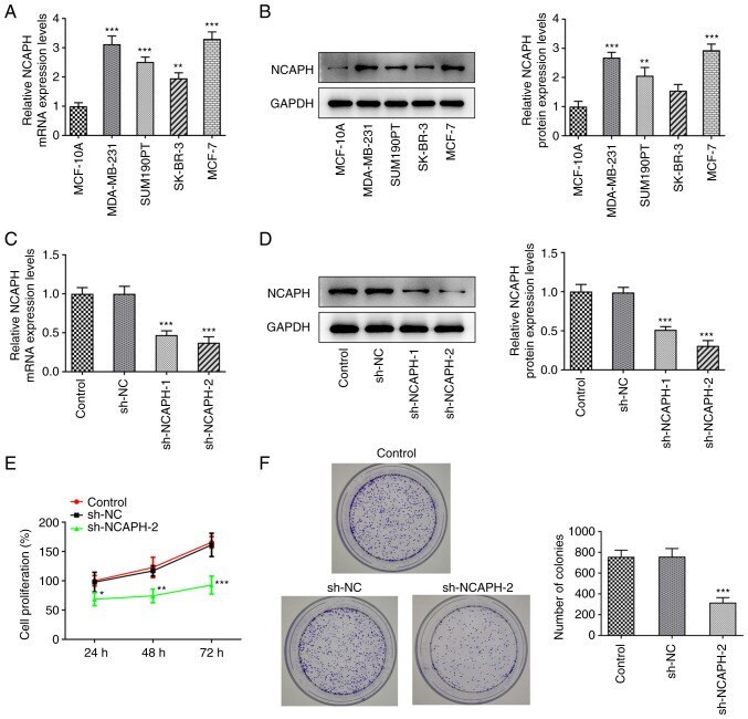
- Figure 1. NCAPH knockdown attenuates breast cancer cell proliferation. mRNA and protein expression levels of NCAPH in MCF-10A cells and four breast cancer cell lines were determined via (A) RT-qPCR and (B) western blotting; respectively. MCF-7 cells were transfected with sh-NCAPH-1/2 and the transfection efficiency was assessed using (C) RT-qPCR and (D) western blotting; respectively. Effect of NCAPH knockdown on cell proliferation was determined using (E) the Cell Counting Kit-8 and (F) colony formation assays. Magnification, x10. *P
- Submitted by
- Invitrogen Antibodies (provider)
- Main image
- Experimental details
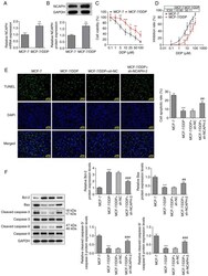
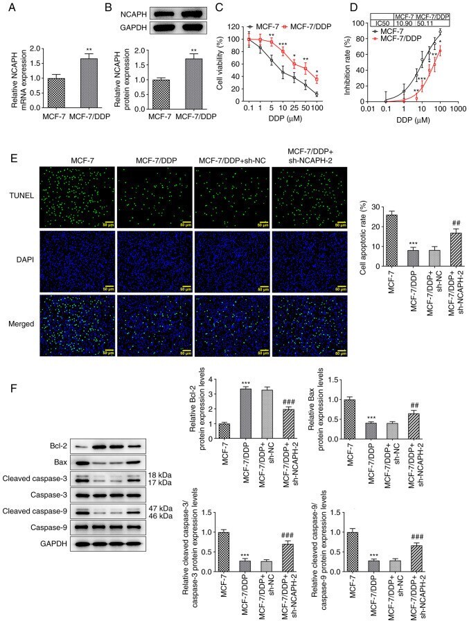
- Figure 3. NCAPH silencing alleviates the resistance of breast cancer cells to DDP. mRNA and protein expression levels of NCAPH in MCF-7 and MCF-7/DDP cells were determined via (A) RT-qPCR and (B) western blotting respectively. (C) Effect of different concentrations of DDP on MCF-7 and MCF-7/DDP cell viability was assessed using the Cell Counting Kit-8 assay. (D) IC 50 values of MCF-7 and MCF-7/DDP cells. (E) Cell apoptotic rate in each group was assessed using the TUNEL assay. Magnification, x200. (F) Protein expression levels of apoptosis-related proteins were detected using western blotting. *P
- Submitted by
- Invitrogen Antibodies (provider)
- Main image
- Experimental details
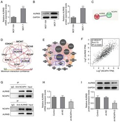
- Figure 4. NCAPH knockdown downregulates AURKB. mRNA and protein expression levels of AURKB in MCF-10A and MCF-7 cells were detected via (A) RT-qPCR and (B) western blotting, respectively. (C) Search Tool for the Retrieval of Interacting Genes/Proteins, (D) HumanBase and (E) GeneMANIA databases were used to predict the association between NCAPH and AURKB. (F) Bioinformatics analysis in the Gene Expression Profiling Interactive Analysis database predicted that NCAPH was positively associated with AURKB. P0 indicates a positive correlation and the closer R 2 is to 1, the more relevant the correlation between NCAPH and AURKB is. (G) Co-immunoprecipitation assay was performed to verify the association between NCAPH and AURKB. mRNA and protein expression levels of AURKB in transfected MCF-7 cells were determined via (H) RT-qPCR and (I) western blotting respectively. **P