Explore
 Validate
 Learn
Learn Western blot
 Immunocytochemistry
Antibody data
- Antibody Data
- Antigen structure
- References [3]
- Comments [0]
- Validations
- Western blot [1]
- Immunocytochemistry [1]
- Immunohistochemistry [1]
Submit
Validation data
Reference
Comment
Report error
- Product number
- HPA003008 - Provider product page

- Provider
- Atlas Antibodies
- Proper citation
- Atlas Antibodies Cat#HPA003008, RRID:AB_1078301
- Product name
- Anti-NCAPH
- Antibody type
- Polyclonal
- Description
- Polyclonal Antibody against Human NCAPH, Gene description: non-SMC condensin I complex, subunit H, Alternative Gene Names: BRRN1, CAP-H, hCAP-H, Validated applications: ICC, IHC, WB, Uniprot ID: Q15003, Storage: Store at +4°C for short term storage. Long time storage is recommended at -20°C.
- Reactivity
- Human
- Host
- Rabbit
- Conjugate
- Unconjugated
- Isotype
- IgG
- Vial size
- 100 µl
- Concentration
- 0.1 mg/ml
- Storage
- Store at +4°C for short term storage. Long time storage is recommended at -20°C.
- Handling
- The antibody solution should be gently mixed before use.
Submitted references Identifying genes as potential prognostic indicators in patients with serous ovarian cancer resistant to carboplatin using integrated bioinformatics analysis
A novel role for the condensin II complex in cellular senescence
Human SMC2 Protein, a Core Subunit of Human Condensin Complex, Is a Novel Transcriptional Target of the WNT Signaling Pathway and a New Therapeutic Target
Zhan S, Liu B, Linghu H
Oncology Reports 2018
Oncology Reports 2018
A novel role for the condensin II complex in cellular senescence
Yokoyama Y, Zhu H, Zhang R, Noma K
Cell Cycle 2015;14(13):2160-2170
Cell Cycle 2015;14(13):2160-2170
Human SMC2 Protein, a Core Subunit of Human Condensin Complex, Is a Novel Transcriptional Target of the WNT Signaling Pathway and a New Therapeutic Target
Dávalos V, Súarez-López L, Castaño J, Messent A, Abasolo I, Fernandez Y, Guerra-Moreno A, Espín E, Armengol M, Musulen E, Ariza A, Sayós J, Arango D, Schwartz S
Journal of Biological Chemistry 2012;287(52):43472-43481
Journal of Biological Chemistry 2012;287(52):43472-43481
No comments: Submit comment
Enhanced validation
- Submitted by
- Atlas Antibodies (provider)
- Enhanced method
- Genetic validation
- Main image
- Experimental details
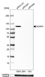
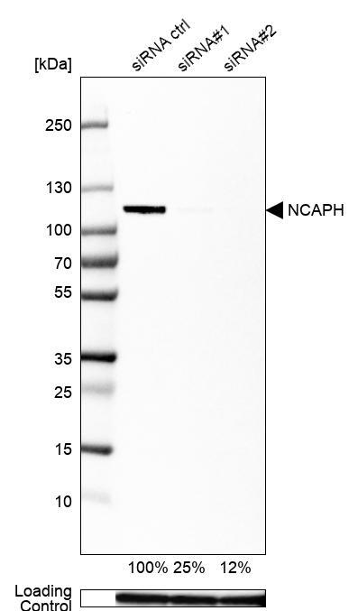
- Western blot analysis in U2OS cells transfected with control siRNA, target specific siRNA probe #1 and #2, using Anti-NCAPH antibody. Remaining relative intensity is presented. Loading control: Anti-GAPDH.
- Sample type
- Human
- Protocol
- Protocol
Supportive validation
- Submitted by
- Atlas Antibodies (provider)
- Main image

- Experimental details
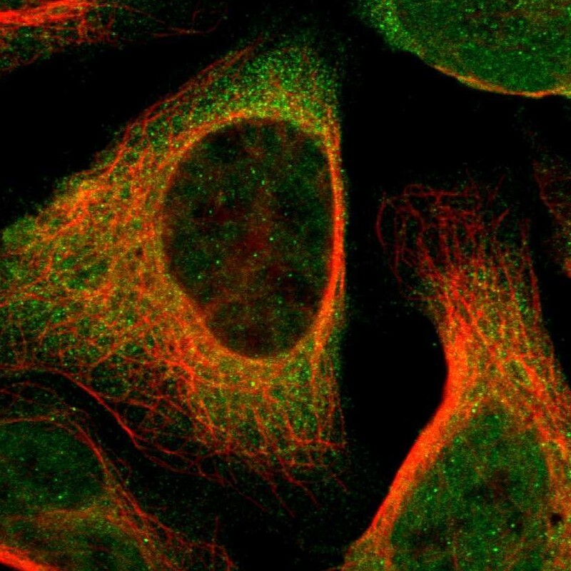
- Immunofluorescent staining of human cell line U-2 OS shows localization to nucleus & cytosol.
- Sample type
- Human
Supportive validation
- Submitted by
- Atlas Antibodies (provider)
- Enhanced method
- Orthogonal validation
- Main image
- Experimental details
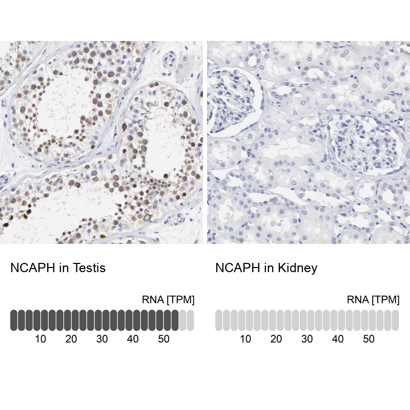
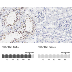
- Immunohistochemistry analysis in human testis and kidney tissues using HPA003008 antibody. Corresponding NCAPH RNA-seq data are presented for the same tissues.
- Sample type
- Human
- Protocol
- Protocol