 Explore
 Validate
 Learn
Learn Western blot
Antibody data
- Antibody Data
- Antigen structure
- References [0]
- Comments [0]
- Validations
- Western blot [1]
- Immunocytochemistry [1]
- Immunohistochemistry [1]
- Flow cytometry [1]
Submit
Validation data
Reference
Comment
Report error
- Product number
- TA329066 - Provider product page

- Provider
- OriGene
- Product name
- Rabbit polyclonal Anti-VPAC1 (extracellular)
- Antibody type
- Polyclonal
- Description
- Rabbit polyclonal Anti-VPAC1 (extracellular)
- Host
- Rabbit
- Conjugate
- Unconjugated
- Epitope
- VIPR1
- Antibody clone number
- NULL
- Vial size
- 200 µl
- Concentration
- NULL
No comments: Submit comment
Supportive validation
- Submitted by
- OriGene (provider)
- Main image
- Experimental details
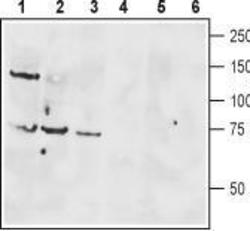
- Western blot analysis of rat brain lysate (lanes 1 and 4), mouse brain membranes (lanes 2 and 5) and human Jurkat T cell leukemia cell lysate (lanes 3 and 6): 1-3. Anti-VPAC1 (extracellular) antibody, (1:400). 4-6. Anti-VPAC1 (extracellular) antibody, preincubated with the control peptide antigen.
- Validation comment
- WB

Supportive validation
- Submitted by
- OriGene (provider)
- Main image
- Experimental details
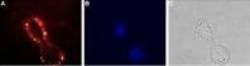
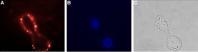
- Expression of VPAC1 in human HT-29 cells. Immunocytochemical staining of live intact human HT-29 colorectal adenocarcinoma cells. A. Extracellular staining of live cells with Anti-VPAC1 (extracellular) antibody, (1:25), followed by goat anti-rabbit-AlexaFluor-594 secondary antibody (red). B. Cell nuclei were visualized using Hoechst 33342 (blue). C. Live view of the cells.
- Validation comment
- IF
Supportive validation
- Submitted by
- OriGene (provider)
- Main image
- Experimental details
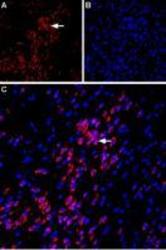
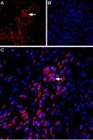
- Expression of VPAC1 in rat amygdala. Immunohistochemical staining of immersion-fixed, free floating rat brain frozen sections using Anti-VPAC1 (extracellular) antibody, (1:100). A. VPAC1 staining (red) is apparent in basolateral amygdala neurons (horizontal arrow). B. Cell nuclei in the same section are stained with DAPI (Blue). C. Merge of the two images.
- Validation comment
- IHC
Supportive validation
- Submitted by
- OriGene (provider)
- Main image
- Experimental details
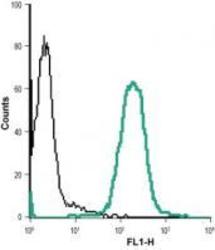
- Indirect flow cytometry analysis of live intact Jurkat (human T cell leukemia cells) cell line: black line Cells + goat-anti-rabbit-DyLight-488. Green line Cells + Anti-VPAC1 (extracellular) antibody , (1:20) + goat-anti-rabbit-DyLight-488.
- Validation comment
- FC
