Explore
 Validate
 Learn
Learn Western blot
Antibody data
- Antibody Data
- Antigen structure
- References [0]
- Comments [0]
- Validations
- Western blot [1]
- Immunocytochemistry [1]
- Immunohistochemistry [1]
- Flow cytometry [1]
Submit
Validation data
Reference
Comment
Report error
- Product number
- AVR-001-200UL - Provider product page

- Provider
- Invitrogen Antibodies
- Product name
- VPAC1 (VIPR1) (extracellular) Polyclonal Antibody
- Antibody type
- Polyclonal
- Antigen
- Other
- Reactivity
- Human, Mouse, Rat
- Host
- Rabbit
- Isotype
- IgG
- Vial size
- 200 µL
- Concentration
- 0.8 mg/mL
- Storage
- -20° C, Avoid Freeze/Thaw Cycles
No comments: Submit comment
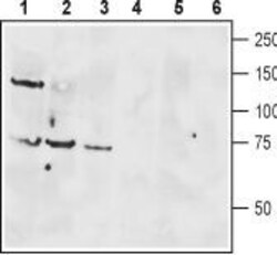
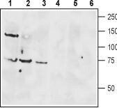
Supportive validation
- Submitted by
- Invitrogen Antibodies (provider)
- Main image
- Experimental details
- Western blot analysis of rat brain lysate (lanes 1 and 4), mouse brain membranes (lanes 2 and 5) and human Jurkat T cell leukemia cell lysate (lanes 3 and 6): - 1-3. Anti-VPAC1 (VIPR1) (extracellular) Antibody (#AVR-001), (1:400).4-6. Anti-VPAC1 (VIPR1) (extracellular) Antibody , preincubated with VPAC1/VIPR1 (extracellular) Blocking Peptide (#BLP-VR001).
Supportive validation
- Submitted by
- Invitrogen Antibodies (provider)
- Main image
- Experimental details
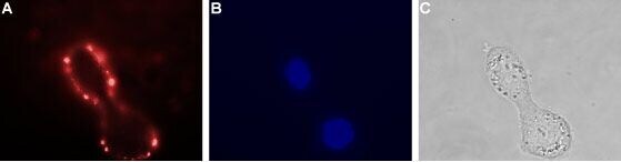
- Expression of VPAC1 in human HT-29 cells - Cell surface detection of VPAC1 in live intact human HT-29 colorectal adenocarcinoma cells. A. Extracellular staining of live cells with Anti-VPAC1 (VIPR1) (extracellular) Antibody (#AVR-001), (1:25), followed by goat Anti-rabbit-AlexaFluor-594 secondary Antibody (red). B. Cell nuclei were visualized using Hoechst 33342 (blue). C. Live view of the cells.
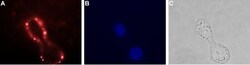
Supportive validation
- Submitted by
- Invitrogen Antibodies (provider)
- Main image
- Experimental details
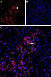
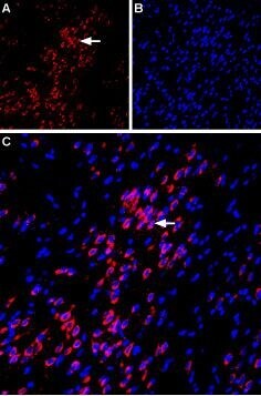
- Expression of VPAC1 in rat amygdala - Immunohistochemical staining of immersion-fixed, free floating rat brain frozen sections using Anti-VPAC1 (VIPR1) (extracellular) Antibody (#AVR-001), (1:100). A. VPAC1 staining (red) is apparent in basolateral amygdala neurons (horizontal arrow). B. Cell nuclei in the same section are stained with DAPI (Blue). C. Merge of the two images.
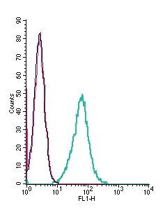
Supportive validation
- Submitted by
- Invitrogen Antibodies (provider)
- Main image

- Experimental details
- Cell surface detection of VPAC1 by indirect flow cytometry in live intact mouse J774 macrophage cells: - (black line) cells. (red) Cells + goat- Anti-rabbit-FITC. (green) Cells + Anti-VPAC1 (VIPR1) (extracellular) Antibody (#AVR-001), (2.5μg) + goat- Anti-rabbit-FITC.