Explore
 Validate
 Learn
Learn Western blot
Antibody data
- Antibody Data
- Antigen structure
- References [0]
- Comments [0]
- Validations
- Western blot [2]
- Immunocytochemistry [1]
Submit
Validation data
Reference
Comment
Report error
- Product number
- AF3917 - Provider product page

- Provider
- R&D Systems
- Product name
- Human MafF Antibody
- Antibody type
- Polyclonal
- Description
- Antigen Affinity-purified. Detects human MafF in direct ELISAs and Western blots. In Western blots, less than 1% cross-reactivity with recombinant human (rh) MafG and rhMafK is observed.
- Reactivity
- Human
- Host
- Goat
- Conjugate
- Unconjugated
- Antigen sequence
Q9ULX9- Isotype
- IgG
- Vial size
- 100 ug
- Concentration
- LYOPH
- Storage
- Use a manual defrost freezer and avoid repeated freeze-thaw cycles. 12 months from date of receipt, -20 to -70 °C as supplied. 1 month, 2 to 8 °C under sterile conditions after reconstitution. 6 months, -20 to -70 °C under sterile conditions after reconstitution.
No comments: Submit comment
Supportive validation
- Submitted by
- R&D Systems (provider)
- Main image

- Experimental details
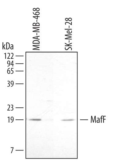
- Detection of Human MafF by Western Blot. Western blot shows lysates of MDA-MB-468 human breast cancer cell line and SK-Mel-28 human malignant melanoma cell line. PVDF membrane was probed with 2 µg/mL Goat Anti-Human MafF Antigen Affinity-purified Polyclonal Antibody (Catalog # AF3917) followed by HRP-conjugated Anti-Goat IgG Secondary Antibody (Catalog # HAF017). A specific band for MafF was detected at approximately 19 kDa (as indicated). This experiment was conducted under reducing conditions and using Immunoblot Buffer Group 1.
- Submitted by
- R&D Systems (provider)
- Main image
- Experimental details
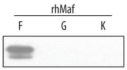
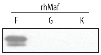
- Detection of Human MafF by Western Blot. Western blot shows recombinant human (rh) MafF, MafG, and MafK (2 ng/lane). PVDF membrane was probed with 2 µg/mL Goat Anti-Human MafF Antigen Affinity-purified Polyclonal Antibody (Catalog # AF3917) followed by HRP-conjugated Anti-Goat IgG Secondary Antibody (Catalog # HAF017). A specific band for MafF was detected at approxi-mately 19 kDa (as indicated). This experiment was conducted under reducing conditions and using Immunoblot Buffer Group 1.
Supportive validation
- Submitted by
- R&D Systems (provider)
- Main image

- Experimental details
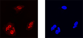
- MafF in HepG2 Human Cell Line. MafF was detected in immersion fixed HepG2 human hepatocellular carcinoma cell line using Goat Anti-Human MafF Antigen Affinity-purified Polyclonal Antibody (Catalog # AF3917) at 15 µg/mL for 3 hours at room temperature. Cells were stained using the NorthernLights™ 557-conjugated Anti-Goat IgG Secondary Antibody (left panel, red; Catalog # NL001) and counterstained with DAPI (right panel, blue). Specific staining was localized to nuclei. View our protocol for Fluorescent ICC Staining of Cells on Coverslips.