 Explore
 Validate
 Learn
Learn Western blot
 Immunocytochemistry
Antibody data
- Antibody Data
- Antigen structure
- References [4]
- Comments [0]
- Validations
- Immunocytochemistry [1]
Submit
Validation data
Reference
Comment
Report error
- Product number
- HPA003363 - Provider product page

- Provider
- Atlas Antibodies
- Proper citation
- Atlas Antibodies Cat#HPA003363, RRID:AB_1078831
- Product name
- Anti-FBXO44
- Antibody type
- Polyclonal
- Description
- Polyclonal Antibody against Human FBXO44, Gene description: F-box protein 44, Alternative Gene Names: FBG3, FBX30, Fbx44, Fbxo6a, MGC14140, Validated applications: WB, IHC, ICC, Uniprot ID: Q9H4M3, Storage: Store at +4°C for short term storage. Long time storage is recommended at -20°C.
- Reactivity
- Human
- Host
- Rabbit
- Conjugate
- Unconjugated
- Isotype
- IgG
- Vial size
- 100 µl
- Concentration
- 0.4 mg/ml
- Storage
- Store at +4°C for short term storage. Long time storage is recommended at -20°C.
- Handling
- The antibody solution should be gently mixed before use.
Submitted references The F-box-only protein 44 regulates pregnane X receptor protein level by ubiquitination and degradation
FBXO44 promotes DNA replication-coupled repetitive element silencing in cancer cells
FBXO44-Mediated Degradation of RGS2 Protein Uniquely Depends on a Cullin 4B/DDB1 Complex
The F-box Protein FBXO44 Mediates BRCA1 Ubiquitination and Degradation
Florke Gee R, Huber A, Wu J, Bajpai R, Loughran A, Pruett-Miller S, Chen T
Acta Pharmaceutica Sinica B 2023;13(11):4523-4534
Acta Pharmaceutica Sinica B 2023;13(11):4523-4534
FBXO44 promotes DNA replication-coupled repetitive element silencing in cancer cells
Shen J, Qiu Z, Wu Q, Finlay D, Garcia G, Sun D, Rantala J, Barshop W, Hope J, Gimple R, Sangfelt O, Bradley L, Wohlschlegel J, Rich J, Spruck C
Cell 2021;184(2):352-369.e23
Cell 2021;184(2):352-369.e23
FBXO44-Mediated Degradation of RGS2 Protein Uniquely Depends on a Cullin 4B/DDB1 Complex
Porter J, Sjögren B, Swaney S, Neubig R
PLOS ONE 2015;10(5):e0123581
PLOS ONE 2015;10(5):e0123581
The F-box Protein FBXO44 Mediates BRCA1 Ubiquitination and Degradation
Lu Y, Li J, Cheng D, Parameswaran B, Zhang S, Jiang Z, Yew P, Peng J, Ye Q, Hu Y
Journal of Biological Chemistry 2012;287(49):41014-41022
Journal of Biological Chemistry 2012;287(49):41014-41022
No comments: Submit comment
Supportive validation
- Submitted by
- Atlas Antibodies (provider)
- Main image
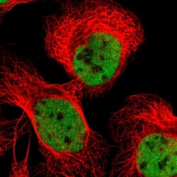
- Experimental details
- Immunofluorescent staining of human cell line U-2 OS shows localization to nucleoplasm.
- Sample type
- Human
