Explore
 Validate
 Learn
Learn Western blot
Antibody data
- Antibody Data
- Antigen structure
- References [0]
- Comments [0]
- Validations
- Western blot [3]
- Immunocytochemistry [1]
- Flow cytometry [1]
Submit
Validation data
Reference
Comment
Report error
- Product number
- ABIN2508617 - Provider product page

- Provider
- antibodies-online
- Product name
- anti-Interleukin 7 (IL7) antibody
- Antibody type
- Polyclonal
- Antigen
- Other
- Description
- Produced from sera of rabbits pre-immunized with highly pure recombinant Human IL-7. Anti-Human IL-7 specific antibody was purified by affinity chromatography employing immobilized Human IL-7 matrix.
- Reactivity
- Human
- Host
- Rabbit
- Vial size
- 100 μg
- Storage
- -20°C
No comments: Submit comment
Supportive validation
- Submitted by
- antibodies-online (provider)
- Main image
- Experimental details
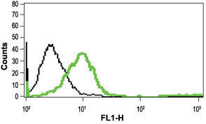
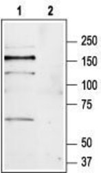
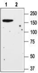
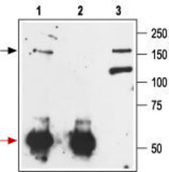
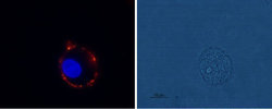
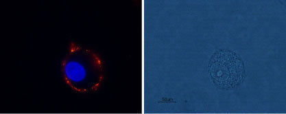
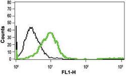
- Western blot analysis of rat brain lysate: 1. Anti-mGluR1 (extracellular) antibody (ABIN2511221), (1:200). 2. Anti-mGluR1 (extracellular) antibody, preincubated with the control peptide antigen. Western blot analysis of mouse brain lysate: 1. Anti-mGluR1 (extracellular) antibody (ABIN2511221), (1:200). 2. Anti-mGluR1 (extracellular) antibody, preincubated with the control peptide antigen. Immunoprecipitation of rat brain lysate: 1. Cell lysate + protein A beads + Anti-mGluR1 (extracellular) antibody (ABIN2511221). 2. Cell lysate + protein A beads + pre-immune rabbit serum. 3. Cell lysate. Expression of mGluR1 in rat C6 glioma cells Immunocytochemical staining of live intact rat C6 glioma cells using Anti-mGluR1 (extracellular) antibody (ABIN2511221), (1:100), followed by goat-anti-rabbit-AlexaFluor-555 secondary antibody (red). Nuclei were stained with Hoechst 33342 (blue). Indirect flow cytometry analysis in live intact Jurkat cells: Black: Unstained cells + goat-anti-rabbit-FITC. Green: Cells + Anti-mGluR1 (extracellular) antibody (ABIN2511221), (10 μg) + goat-anti-rabbit-FITC.

- Submitted by
- antibodies-online (provider)
- Main image
- Experimental details
- Western blot analysis of rat brain lysate: 1. Anti-mGluR1 (extracellular) antibody (ABIN2511221), (1:200). 2. Anti-mGluR1 (extracellular) antibody, preincubated with the control peptide antigen. Western blot analysis of mouse brain lysate: 1. Anti-mGluR1 (extracellular) antibody (ABIN2511221), (1:200). 2. Anti-mGluR1 (extracellular) antibody, preincubated with the control peptide antigen. Immunoprecipitation of rat brain lysate: 1. Cell lysate + protein A beads + Anti-mGluR1 (extracellular) antibody (ABIN2511221). 2. Cell lysate + protein A beads + pre-immune rabbit serum. 3. Cell lysate. Expression of mGluR1 in rat C6 glioma cells Immunocytochemical staining of live intact rat C6 glioma cells using Anti-mGluR1 (extracellular) antibody (ABIN2511221), (1:100), followed by goat-anti-rabbit-AlexaFluor-555 secondary antibody (red). Nuclei were stained with Hoechst 33342 (blue). Indirect flow cytometry analysis in live intact Jurkat cells: Black: Unstained cells + goat-anti-rabbit-FITC. Green: Cells + Anti-mGluR1 (extracellular) antibody (ABIN2511221), (10 μg) + goat-anti-rabbit-FITC.

Supportive validation
- Submitted by
- antibodies-online (provider)
- Main image
- Experimental details
- Image(s): Immunofluorescence
Supportive validation
- Submitted by
- antibodies-online (provider)
- Main image
- Experimental details
- Image(s): Flow cytometry