Explore
 Validate
 Learn
Learn Western blot
 ELISA
ELISA Immunocytochemistry
Antibody data
- Antibody Data
- Antigen structure
- References [11]
- Comments [0]
- Validations
- Immunocytochemistry [2]
- Other assay [8]
Submit
Validation data
Reference
Comment
Report error
- Product number
- 37-3000 - Provider product page

- Provider
- Invitrogen Antibodies
- Product name
- RPL11 Monoclonal Antibody (3A4A7)
- Antibody type
- Monoclonal
- Antigen
- Other
- Reactivity
- Human
- Host
- Mouse
- Isotype
- IgG
- Antibody clone number
- 3A4A7
- Vial size
- 100 μg
- Concentration
- 0.5 mg/mL
- Storage
- -20°C
Submitted references Adaptive translational reprogramming of metabolism limits the response to targeted therapy in BRAF(V600) melanoma.
The exon-junction complex helicase eIF4A3 controls cell fate via coordinated regulation of ribosome biogenesis and translational output.
Ribosomal Protein L15 is involved in Colon Carcinogenesis.
Direct relationship between the level of p53 stabilization induced by rRNA synthesis-inhibiting drugs and the cell ribosome biogenesis rate.
Acrolein preferentially damages nucleolus eliciting ribosomal stress and apoptosis in human cancer cells.
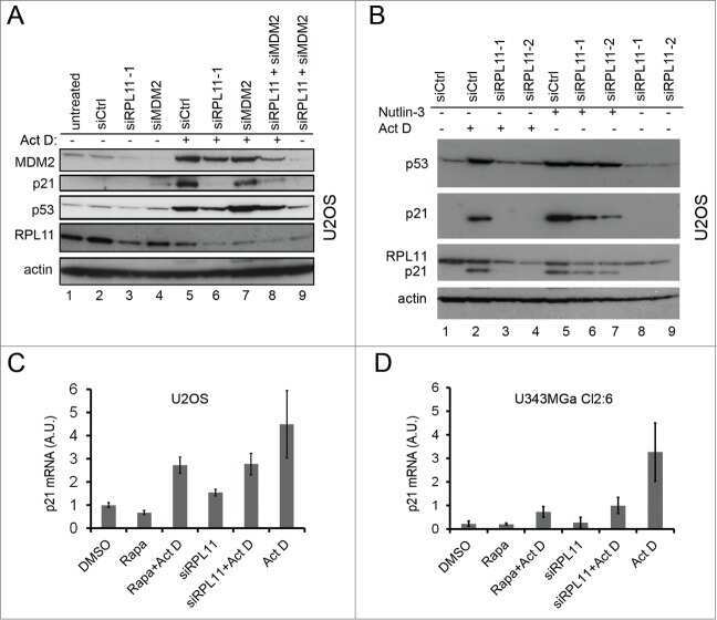
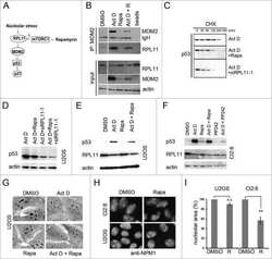
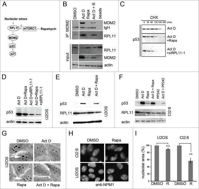
mTOR inhibitors blunt the p53 response to nucleolar stress by regulating RPL11 and MDM2 levels.
Growth inhibitory effects of large subunit ribosomal proteins in melanoma.
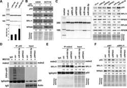
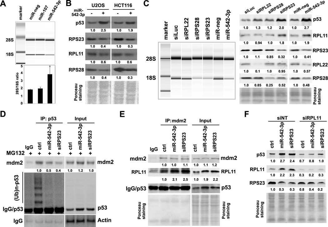
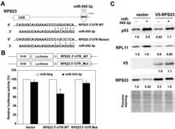
p53 is positively regulated by miR-542-3p.
5S ribosomal RNA is an essential component of a nascent ribosomal precursor complex that regulates the Hdm2-p53 checkpoint.
Ribosomal protein S6 interacts with the latency-associated nuclear antigen of Kaposi's sarcoma-associated herpesvirus.
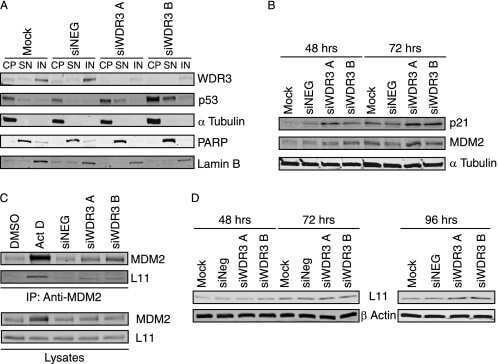
Ribosomal 18 S RNA processing by the IGF-I-responsive WDR3 protein is integrated with p53 function in cancer cell proliferation.
Smith LK, Parmenter T, Kleinschmidt M, Kusnadi EP, Kang J, Martin CA, Lau P, Patel R, Lorent J, Papadopoli D, Trigos A, Ward T, Rao AD, Lelliott EJ, Sheppard KE, Goode D, Hicks RJ, Tiganis T, Simpson KJ, Larsson O, Blythe B, Cullinane C, Wickramasinghe VO, Pearson RB, McArthur GA
Nature communications 2022 Mar 1;13(1):1100
Nature communications 2022 Mar 1;13(1):1100
The exon-junction complex helicase eIF4A3 controls cell fate via coordinated regulation of ribosome biogenesis and translational output.
Kanellis DC, Espinoza JA, Zisi A, Sakkas E, Bartkova J, Katsori AM, Boström J, Dyrskjøt L, Broholm H, Altun M, Elsässer SJ, Lindström MS, Bartek J
Science advances 2021 Aug;7(32)
Science advances 2021 Aug;7(32)
Ribosomal Protein L15 is involved in Colon Carcinogenesis.
Dong Z, Jiang H, Liang S, Wang Y, Jiang W, Zhu C
International journal of medical sciences 2019;16(8):1132-1141
International journal of medical sciences 2019;16(8):1132-1141
Direct relationship between the level of p53 stabilization induced by rRNA synthesis-inhibiting drugs and the cell ribosome biogenesis rate.
Scala F, Brighenti E, Govoni M, Imbrogno E, Fornari F, Treré D, Montanaro L, Derenzini M
Oncogene 2016 Feb 25;35(8):977-89
Oncogene 2016 Feb 25;35(8):977-89
Acrolein preferentially damages nucleolus eliciting ribosomal stress and apoptosis in human cancer cells.
Wang HT, Chen TY, Weng CW, Yang CH, Tang MS
Oncotarget 2016 Dec 6;7(49):80450-80464
Oncotarget 2016 Dec 6;7(49):80450-80464
mTOR inhibitors blunt the p53 response to nucleolar stress by regulating RPL11 and MDM2 levels.
Goudarzi KM, Nistér M, Lindström MS
Cancer biology & therapy 2014;15(11):1499-514
Cancer biology & therapy 2014;15(11):1499-514
Growth inhibitory effects of large subunit ribosomal proteins in melanoma.
Kardos GR, Dai MS, Robertson GP
Pigment cell & melanoma research 2014 Sep;27(5):801-12
Pigment cell & melanoma research 2014 Sep;27(5):801-12
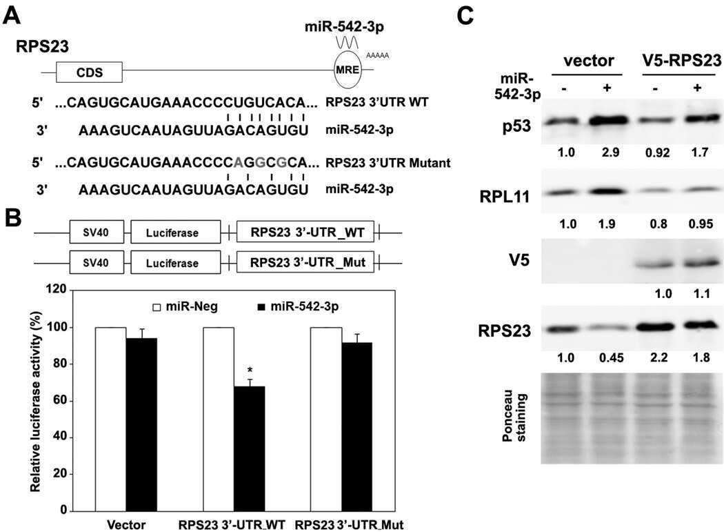
p53 is positively regulated by miR-542-3p.
Wang Y, Huang JW, Castella M, Huntsman DG, Taniguchi T
Cancer research 2014 Jun 15;74(12):3218-27
Cancer research 2014 Jun 15;74(12):3218-27
5S ribosomal RNA is an essential component of a nascent ribosomal precursor complex that regulates the Hdm2-p53 checkpoint.
Donati G, Peddigari S, Mercer CA, Thomas G
Cell reports 2013 Jul 11;4(1):87-98
Cell reports 2013 Jul 11;4(1):87-98
Ribosomal protein S6 interacts with the latency-associated nuclear antigen of Kaposi's sarcoma-associated herpesvirus.
Chen W, Dittmer DP
Journal of virology 2011 Sep;85(18):9495-505
Journal of virology 2011 Sep;85(18):9495-505
Ribosomal 18 S RNA processing by the IGF-I-responsive WDR3 protein is integrated with p53 function in cancer cell proliferation.
McMahon M, Ayllón V, Panov KI, O'Connor R
The Journal of biological chemistry 2010 Jun 11;285(24):18309-18
The Journal of biological chemistry 2010 Jun 11;285(24):18309-18
No comments: Submit comment
Supportive validation
- Submitted by
- Invitrogen Antibodies (provider)
- Main image

- Experimental details
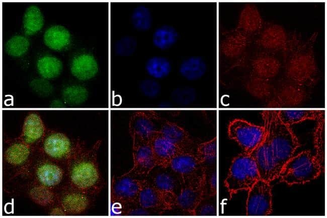
- Immunofluorescence analysis of L11 was performed using 70% confluent log phase U-2 OS cells treated with doxorubicin (2 µg/mL) for 24 hours. The cells were fixed with 4% paraformaldehyde for 10 minutes, permeabilized with 0.1% Triton™ X-100 for 10 minutes, and blocked with 1% BSA for 1 hour at room temperature. The cells were labeled with RPL11 (3A4A7) Mouse Monoclonal Antibody (Product # 37-3000) at 2 µg/mL in 0.1% BSA and incubated for 3 hours at room temperature and then labeled with Goat anti-Mouse IgG (H+L) Superclonal™ Secondary Antibody, Alexa Fluor® 488 conjugate (Product # A28175) at a dilution of 1:2000 for 45 minutes at room temperature (Panel a: green). Nuclei (Panel b: blue) were stained with SlowFade® Gold Antifade Mountant with DAPI (Product # S36938). F-actin (Panel c: red) was stained with Alexa Fluor® 555 Rhodamine Phalloidin (Product # R415, 1:300). Panel d represents the merged image showing nuclear localization. Panel e is untreated cell with no signal. Panel f represents control cells with no primary antibody. The images were captured at 60X magnification.
- Submitted by
- Invitrogen Antibodies (provider)
- Main image
- Experimental details
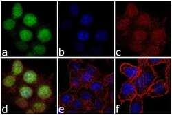
- Immunofluorescence analysis of L11 was performed using 70% confluent log phase U-2 OS cells treated with doxorubicin (2 µg/mL) for 24 hours. The cells were fixed with 4% paraformaldehyde for 10 minutes, permeabilized with 0.1% Triton™ X-100 for 10 minutes, and blocked with 1% BSA for 1 hour at room temperature. The cells were labeled with RPL11 (3A4A7) Mouse Monoclonal Antibody (Product # 37-3000) at 2 µg/mL in 0.1% BSA and incubated for 3 hours at room temperature and then labeled with Goat anti-Mouse IgG (H+L) Superclonal™ Secondary Antibody, Alexa Fluor® 488 conjugate (Product # A28175) at a dilution of 1:2000 for 45 minutes at room temperature (Panel a: green). Nuclei (Panel b: blue) were stained with SlowFade® Gold Antifade Mountant with DAPI (Product # S36938). F-actin (Panel c: red) was stained with Alexa Fluor® 555 Rhodamine Phalloidin (Product # R415, 1:300). Panel d represents the merged image showing nuclear localization. Panel e is untreated cell with no signal. Panel f represents control cells with no primary antibody. The images were captured at 60X magnification.
Supportive validation
- Submitted by
- Invitrogen Antibodies (provider)
- Main image

- Experimental details
- NULL
- Submitted by
- Invitrogen Antibodies (provider)
- Main image
- Experimental details
- NULL
- Submitted by
- Invitrogen Antibodies (provider)
- Main image
- Experimental details
- NULL
- Submitted by
- Invitrogen Antibodies (provider)
- Main image
- Experimental details
- NULL
- Submitted by
- Invitrogen Antibodies (provider)
- Main image
- Experimental details
- NULL
- Submitted by
- Invitrogen Antibodies (provider)
- Main image
- Experimental details
- NULL
- Submitted by
- Invitrogen Antibodies (provider)
- Main image
- Experimental details
- NULL
- Submitted by
- Invitrogen Antibodies (provider)
- Main image
- Experimental details
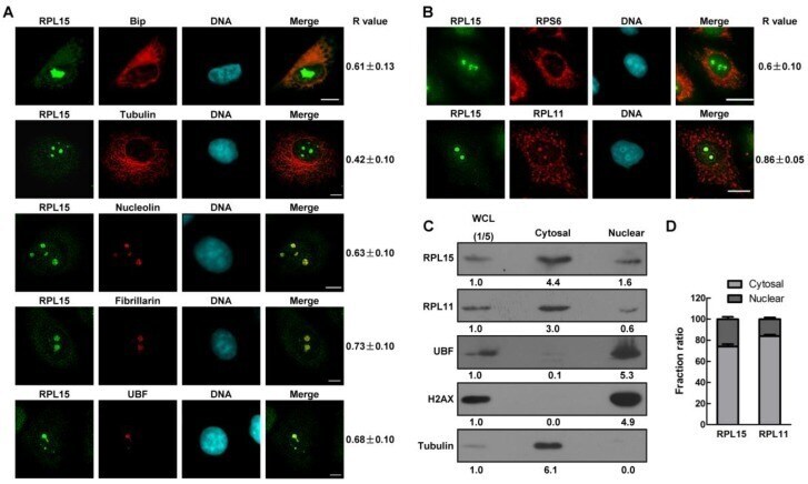
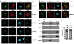
- Figure 1 Characterization of ribosomal protein RPL15. (A) Immunofluorescence analysis of HeLa cells with rabbit alpha-RPL15 and mouse alpha-Bip, anti-alpha-tubulin, alpha-nucleolin, alpha-fibrillarin or alpha-UBF. DNA was visualized by DAPI staining. R values were obtained as described (see Materials and Methods). Scale bars, 10 mum. (B) Immunofluorescence analysis of HeLa cells with rabbit alpha-RPL15 and mouse alpha-RPL11 or mouse alpha-RPS6 and DAPI. Scale bars, 10 mum. R values were obtained as described in (A) . (C) Nuclear or cytoplasmic lysates from HeLa cells were extracted and subjected to immunoblotting by indicated antibodies. The density of each protein in each component was quantitated against the level of WCL (1/5 of total). WCL: whole cell lysate. (D) Histograms represented average proportion of RPL15 or RPL11 in Nuclear or cytoplasmic component relative to WCL in (C). All experiments were performed in triplicate. Data are shown as mean +- standard deviations.