Explore
 Validate
 Learn
Learn Western blot
 Chromatin Immunoprecipitation
Antibody data
- Antibody Data
- Antigen structure
- References [0]
- Comments [0]
- Validations
- Western blot [1]
- Immunohistochemistry [6]
- Other assay [1]
Submit
Validation data
Reference
Comment
Report error
- Product number
- UM500047CF - Provider product page

- Provider
- Invitrogen Antibodies
- Product name
- SOX5 Monoclonal Antibody (UMAB53), UltraMAB™
- Antibody type
- Monoclonal
- Antigen
- Recombinant full-length protein
- Reactivity
- Human
- Host
- Mouse
- Isotype
- IgG
- Antibody clone number
- UMAB53
- Vial size
- 100 µg
- Concentration
- 1 mg/mL
- Storage
- -20° C, Avoid Freeze/Thaw Cycles
No comments: Submit comment
Supportive validation
- Submitted by
- Invitrogen Antibodies (provider)
- Main image
- Experimental details
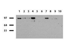
- Western blot of human tissue lysates (15 µg) from 10 different tissues (1: Testis, 2: Omentum, 3: Uterus, 4: Breast, 5: Brain, 6: Thyroid, 7: Colon, 8: Spleen 9: Liver, 10: Ovary). Dilution: 1:500.

Supportive validation
- Submitted by
- Invitrogen Antibodies (provider)
- Main image

- Experimental details
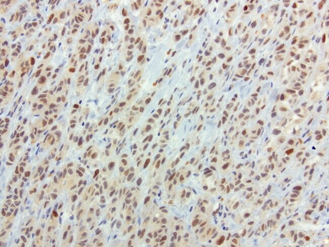
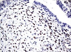
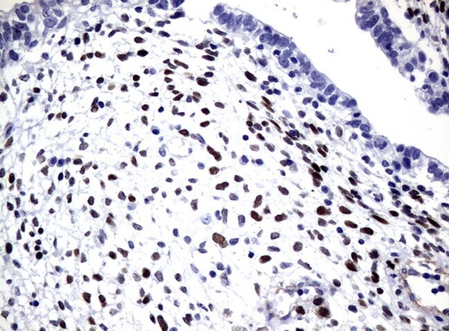
- Immunohistochemical staining of paraffin-embedded human melanoma using anti-SOX5 clone UMAB53 mouse monoclonal antibody (UM500047) at 1:100 with Polink2 Broad HRP DAB detection kit; heat-induced epitope retrieval with GBI Accel pH 8.7 HIER buffer using pressure chamber for 3 minutes at 110°C. Strong nuclear and weak cytoplasmic staining is seen in the tumor cells.
- Submitted by
- Invitrogen Antibodies (provider)
- Main image
- Experimental details
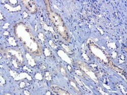
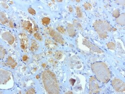
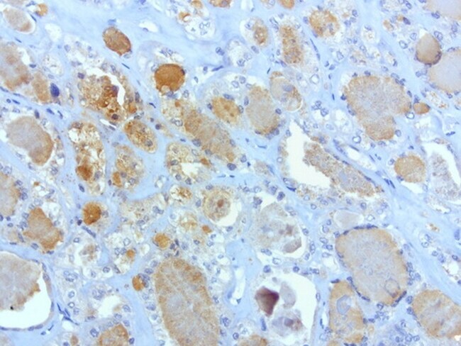
- Immunohistochemical staining of paraffin-embedded human kidney using anti-SOX5 clone UMAB53 mouse monoclonal antibody (UM500047) at 1:100 with Polink2 Broad HRP DAB detection kit; heat-induced epitope retrieval with GBI Accel pH 8.7 HIER buffer using pressure chamber for 3 minutes at 110°C. Weak cytoplasmic staining is seen in the tubule epithelial cells of kidney.

- Submitted by
- Invitrogen Antibodies (provider)
- Main image
- Experimental details
- Immunohistochemical staining of paraffin-embedded human pancreatic cancer using anti-SOX5 clone UMAB53 mouse monoclonal antibody (UM500047) at 1:100 with Polink2 Broad HRP DAB detection kit; heat-induced epitope retrieval with GBI Accel pH 8.7 HIER buffer using pressure chamber for 3 minutes at 110°C. Strong cytoplasmic staining is seen in the tumor cells.
- Submitted by
- Invitrogen Antibodies (provider)
- Main image
- Experimental details
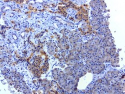
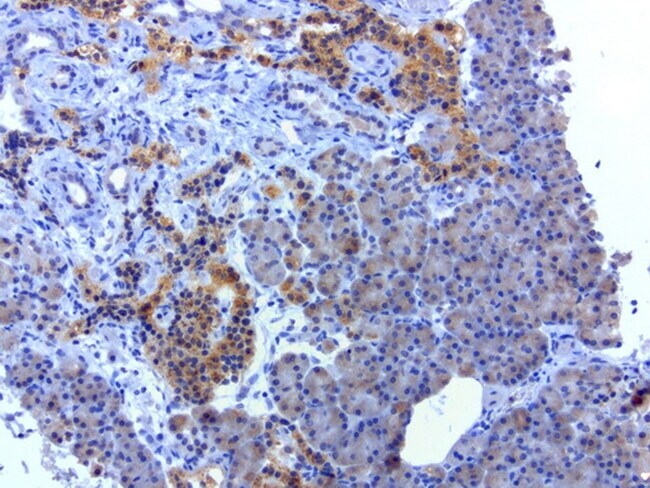
- Immunohistochemical staining of paraffin-embedded human thyroid cancer using anti-SOX5 clone UMAB53 mouse monoclonal antibody (UM500047) at 1:100 with Polink2 Broad HRP DAB detection kit; heat-induced epitope retrieval with GBI Accel pH 8.7 HIER buffer using pressure chamber for 3 minutes at 110°C. Cytoplasmic staining is seen in the tumor cells.
- Submitted by
- Invitrogen Antibodies (provider)
- Main image
- Experimental details
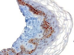
- Immunohistochemical staining of paraffin-embedded human skin using anti-SOX5 clone UMAB53 mouse monoclonal antibody (UM500047) at 1:100 with Polink2 Broad HRP DAB detection kit; heat-induced epitope retrieval with GBI Accel pH 8.7 HIER buffer using pressure chamber for 3 minutes at 110°C. Strong nuclear and cytoplasmic staining is seen in the epidermal cells.

- Submitted by
- Invitrogen Antibodies (provider)
- Main image
- Experimental details
- Immunohistochemical staining of paraffin-embedded human testicular cancer tissue using anti-SOX5 mouse monoclonal antibody. (UM500047; heat-induced epitope retrieval by 10mM citric buffer, pH6.0, 120°C for 3min)
Supportive validation
- Submitted by
- Invitrogen Antibodies (provider)
- Main image
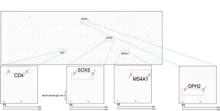
- Experimental details
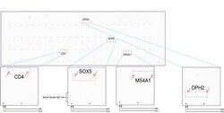
- OriGene overexpression protein microarray chip was immunostained with UltraMAB anti-SOX5 mouse monoclonal antibody (UM500047). The positive reactive proteins are highlighted with two red arrows in the enlarged subarray. All the positive controls spotted in this subarray are also labeled for clarification.