Explore
 Validate
 Learn
Learn Western blot
 Immunohistochemistry
Antibody data
- Antibody Data
- Antigen structure
- References [2]
- Comments [0]
- Validations
- Immunohistochemistry [1]
- Other assay [2]
Submit
Validation data
Reference
Comment
Report error
- Product number
- PA5-31392 - Provider product page

- Provider
- Invitrogen Antibodies
- Product name
- SMOC1 Polyclonal Antibody
- Antibody type
- Polyclonal
- Antigen
- Recombinant full-length protein
- Description
- Recommended positive controls: U87-MG. Predicted reactivity: Mouse (96%), Rat (95%), Xenopus laevis (84%), Bovine (95%). Store product as a concentrated solution. Centrifuge briefly prior to opening the vial.
- Reactivity
- Human
- Host
- Rabbit
- Isotype
- IgG
- Vial size
- 100 μL
- Concentration
- 0.05 mg/mL
- Storage
- Store at 4°C short term. For long term storage, store at -20°C, avoiding freeze/thaw cycles.
Submitted references Pathogenetic Mechanisms Underlying Spinocerebellar Ataxia Type 3 Are Altered in Primary Oligodendrocyte Culture.
The amyloid plaque proteome in early onset Alzheimer's disease and Down syndrome.
Schuster KH, Putka AF, McLoughlin HS
Cells 2022 Aug 22;11(16)
Cells 2022 Aug 22;11(16)
The amyloid plaque proteome in early onset Alzheimer's disease and Down syndrome.
Drummond E, Kavanagh T, Pires G, Marta-Ariza M, Kanshin E, Nayak S, Faustin A, Berdah V, Ueberheide B, Wisniewski T
Acta neuropathologica communications 2022 Apr 13;10(1):53
Acta neuropathologica communications 2022 Apr 13;10(1):53
No comments: Submit comment
Supportive validation
- Submitted by
- Invitrogen Antibodies (provider)
- Main image
- Experimental details
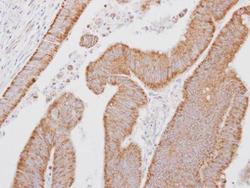
- Immunohistochemical analysis of paraffin-embedded human colon carcinoma, using SMOC1 (Product # PA5-31392) antibody at 1:250 dilution. Antigen Retrieval: EDTA based buffer, pH 8.0, 15 min.

Supportive validation
- Submitted by
- Invitrogen Antibodies (provider)
- Main image
- Experimental details
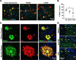
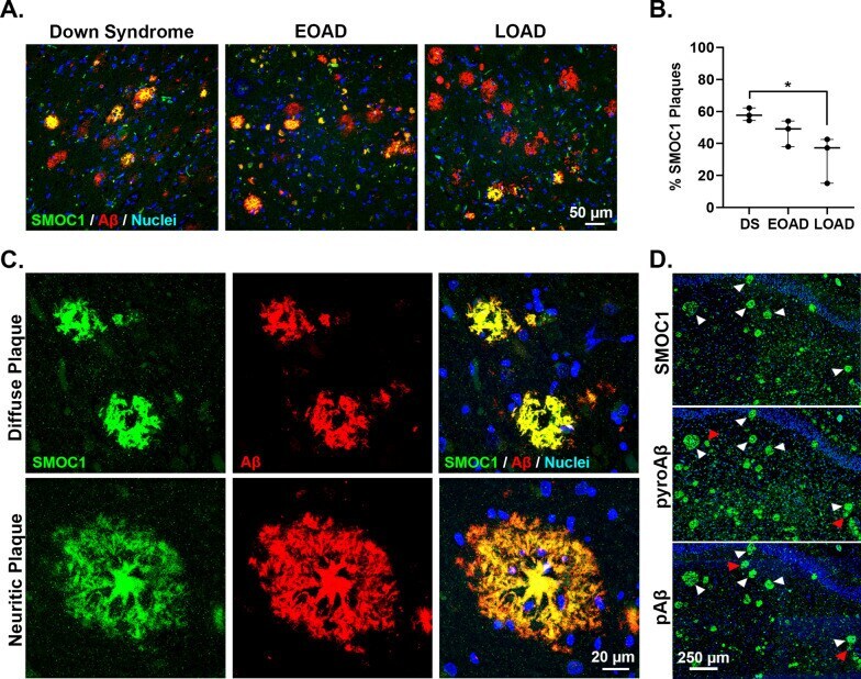
- Validation of SMOC1 as a plaque enriched protein in human brain tissue by immunohistochemistry. A Enrichment of SMOC1 was observed in a sub-population of amyloid plaques in DS, EOAD and LOAD cases. B Plot shows percentage of SMOC1 immunoreactive plaques in the hippocampus of DS, EOAD and LOAD cases (n = 3/group). Results generated by an analysis of 321 +- 47 hippocampal plaques (average +- SEM) in each case. The ratio of SMOC1 positive plaques (immunoreactive for both Abeta and SMOC1) over the total number of amyloid plaques was calculated for each case in DS, EOAD and LOAD. C Representative images of diffuse and neuritic plaques immunolabeled with SMOC1. D Representative images of SMOC1, pyroglutamate Abeta and phosphorylated Abeta immunolabelled plaques in the hippocampus of a representative Down syndrome case. Fluorescent immunohistochemistry was used to identify SMOC1, pyroglutamate Abeta or phosphorylated Abeta immunoreactive plaques on three sequential hippocampal sections from the same case. White arrowheads show SMOC1 immunoreactive amyloid plaques that were also immunoreactive for pyroglutamate Abeta and phosphorylated Abeta species. Red arrowheads show pyroglutamate Abeta and/or phosphorylated Abeta immunoreactive plaques negative for SMOC1. * p < 0.05
- Submitted by
- Invitrogen Antibodies (provider)
- Main image

- Experimental details
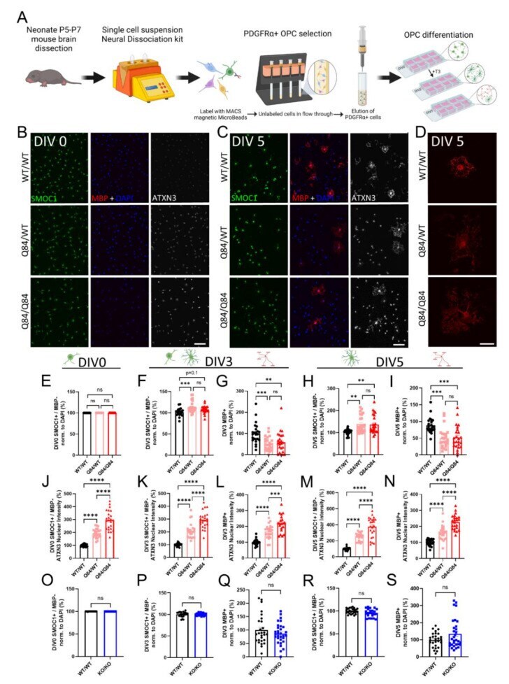
- Figure 1 ATXN3 gain of toxic function, but not loss of function, leads to cell-autonomous oligodendrocyte maturation impairments. ( A ) Schematic of primary OPC isolation and culture. ( B , C ) Representative immunofluorescent images of SMOC1 (green), MBP (red), ATXN3 (white), and DAPI (blue) expression in cultured WT/WT, Q84/WT, and Q84/Q84 oligodendrocytes prior to differentiation (DIV0) ( B ) and after 5 days of differentiation (+T3, DIV5) ( C ). Scale bar, 100 um. ( D ) Representative high magnification images of MBP staining of cultured WT/WT, Q84/WT, and Q84/Q84 oligodendrocytes depict irregular branching in Q84 cells. Scale bar: 25 um. ( E , F , G ) Cell counts of immature oligodendrocytes (SMOC1+/MBP-) at DIV0 ( E ), DIV3 ( F ), and DIV5 ( H ). ( G , I ) Cell counts of mature oligodendrocytes (MBP+) at DIV3 ( G ) and DIV5 ( I ). No differences in cell counts were found at DIV0, however, at DIV3 and DIV5, immature oligodendrocytes were increased and mature oligodendrocytes were significantly decreased in diseased mice. ( J ) Quantification of average ATXN3 nuclear intensity in Q84 OPCs at DIV0. ( K - N ) Quantification of average ATXN3 intensity in Q84 immature and mature oligodendrocyte nuclei at DIV3 ( K , L ) and DIV5 ( M , N ). Oligodendrocytes from diseased mice, regardless of maturation state, show a dose-dependent increase in nuclear ATXN3 accumulation. ( O - S ) Cell counts of OPCs cultured from Atxn3 -KO mice at DIV0 ( O ), of immature and mature oligodendrocy