Explore
 Validate
 Learn
Learn Western blot
 Immunoprecipitation
Antibody data
- Antibody Data
- Antigen structure
- References [0]
- Comments [0]
- Validations
- Western blot [11]
- Immunocytochemistry [2]
- Immunohistochemistry [4]
- Other assay [1]
Submit
Validation data
Reference
Comment
Report error
- Product number
- 21705-1-AP - Provider product page

- Provider
- Invitrogen Antibodies
- Product name
- UQCRC1 Polyclonal Antibody
- Antibody type
- Polyclonal
- Antigen
- Other
- Reactivity
- Human, Mouse, Rat
- Host
- Rabbit
- Isotype
- IgG
- Vial size
- 150 µL
- Concentration
- 0.15 mg/mL
- Storage
- -20°C
No comments: Submit comment
Supportive validation
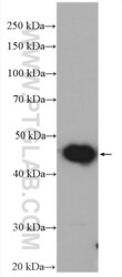
- Submitted by
- Invitrogen Antibodies (provider)
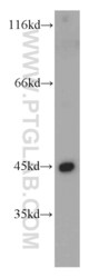
- Main image

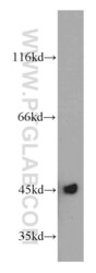
- Experimental details
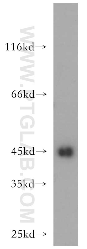
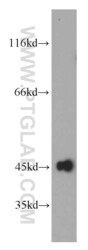
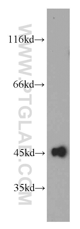
- HeLa cells were subjected to SDS PAGE followed by western blot with 21705-1-AP (UQCRC1 antibody) at dilution of 1:500 incubated at room temperature for 1.5 hours.
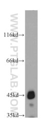
- Submitted by
- Invitrogen Antibodies (provider)
- Main image
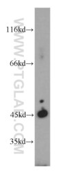
- Experimental details
- MCF7 cells were subjected to SDS PAGE followed by western blot with 21705-1-AP (UQCRC1 antibody) at dilution of 1:500 incubated at room temperature for 1.5 hours.
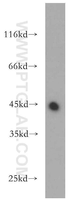
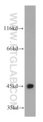
- Submitted by
- Invitrogen Antibodies (provider)
- Main image
- Experimental details
- Mouse liver tissue were subjected to SDS PAGE followed by western blot with 21705-1-AP (UQCRC1 antibody) at dilution of 1:2000 incubated at room temperature for 1.5 hours.

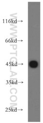
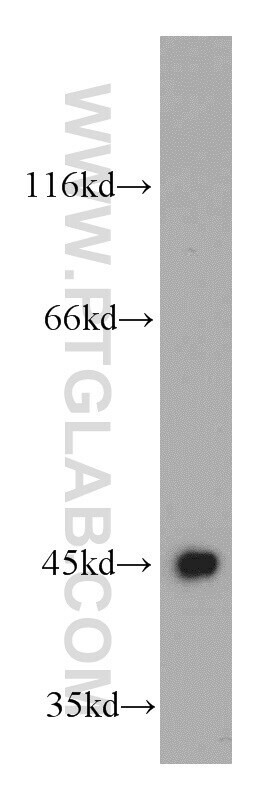
- Submitted by
- Invitrogen Antibodies (provider)
- Main image

- Experimental details
- HepG2 cells were subjected to SDS PAGE followed by western blot with 21705-1-AP (UQCRC1 antibody) at dilution of 1:500 incubated at room temperature for 1.5 hours.
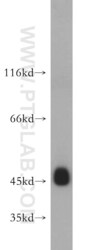
- Submitted by
- Invitrogen Antibodies (provider)
- Main image
- Experimental details
- Human brain tissue were subjected to SDS PAGE followed by western blot with 21705-1-AP (UQCRC1 antibody) at dilution of 1:500 incubated at room temperature for 1.5 hours.

- Submitted by
- Invitrogen Antibodies (provider)
- Main image
- Experimental details
- Human skeletal muscle tissue were subjected to SDS PAGE followed by western blot with 21705-1-AP (UQCRC1 antibody) at dilution of 1:300 incubated at room temperature for 1.5 hours.

- Submitted by
- Invitrogen Antibodies (provider)
- Main image
- Experimental details
- Mouse skeletal muscle tissue were subjected to SDS PAGE followed by western blot with 21705-1-AP (UQCRC1 antibody) at dilution of 1:500 incubated at room temperature for 1.5 hours.

- Submitted by
- Invitrogen Antibodies (provider)
- Main image
- Experimental details
- Human heart tissue were subjected to SDS PAGE followed by western blot with 21705-1-AP (UQCRC1 antibody) at dilution of 1:500 incubated at room temperature for 1.5 hours.

- Submitted by
- Invitrogen Antibodies (provider)
- Main image
- Experimental details
- L02 cells were subjected to SDS PAGE followed by western blot with 21705-1-AP (UQCRC1 antibody) at dilution of 1:500 incubated at room temperature for 1.5 hours.
- Submitted by
- Invitrogen Antibodies (provider)
- Main image
- Experimental details
- HeLa cells were subjected to SDS PAGE followed by western blot with 21705-1-AP (UQCRC1 antibody) at dilution of 1:500 incubated at room temperature for 1.5 hours.

- Submitted by
- Invitrogen Antibodies (provider)
- Main image
- Experimental details
- Mouse large intestine tissue were subjected to SDS PAGE followed by western blot with 21705-1-AP (UQCRC1 antibody) at dilution of 1:500 incubated at room temperature for 1.5 hours.

Supportive validation
- Submitted by
- Invitrogen Antibodies (provider)
- Main image
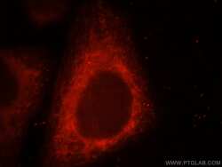
- Experimental details
- Immunofluorescent analysis of HepG2 cells, using UQCRC1 antibody 21705-1-AP at 1:25 dilution and Rhodamine-labeled goat anti-rabbit IGG (red).

- Submitted by
- Invitrogen Antibodies (provider)
- Main image
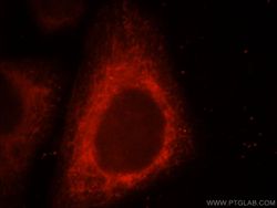
- Experimental details
- Immunofluorescent analysis of HepG2 cells, using UQCRC1 antibody 21705-1-AP at 1:25 dilution and Rhodamine-labeled goat anti-rabbit IGG (red).

Supportive validation
- Submitted by
- Invitrogen Antibodies (provider)
- Main image
- Experimental details
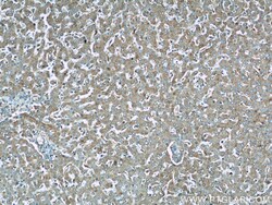
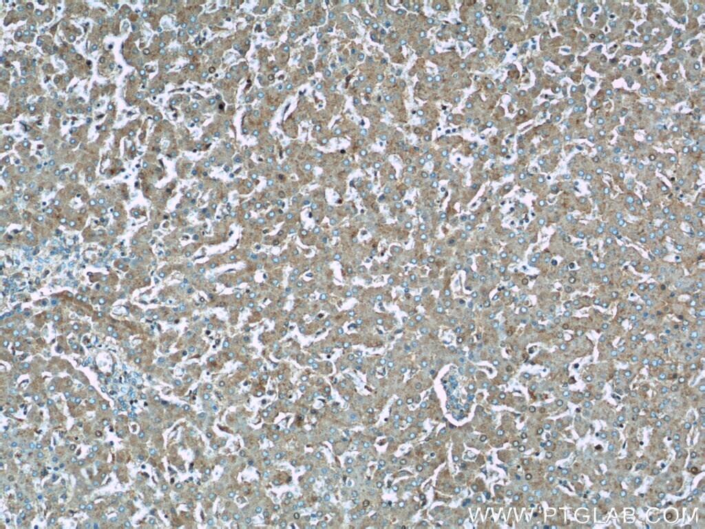
- Immunohistochemistry of paraffin-embedded human liver using 21705-1-AP (UQCRC1 antibody) at dilution of 1:100 (under 10x lens).
- Submitted by
- Invitrogen Antibodies (provider)
- Main image
- Experimental details
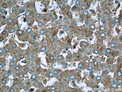
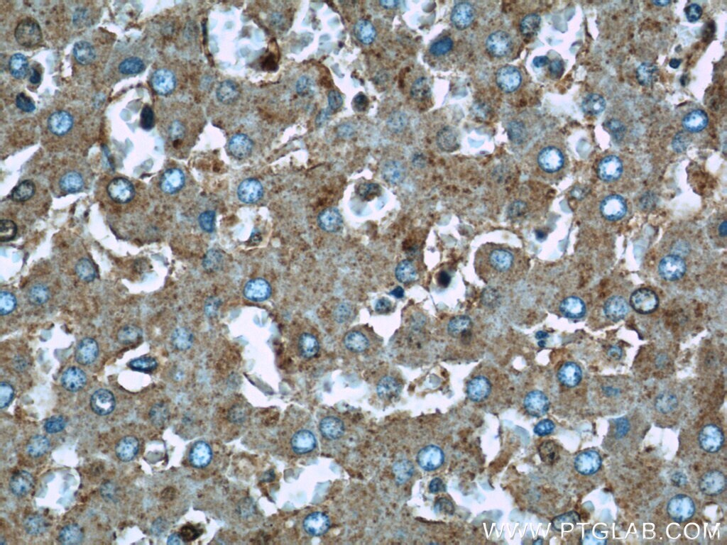
- Immunohistochemistry of paraffin-embedded human liver using 21705-1-AP (UQCRC1 antibody) at dilution of 1:100 (under 40x lens).
- Submitted by
- Invitrogen Antibodies (provider)
- Main image

- Experimental details
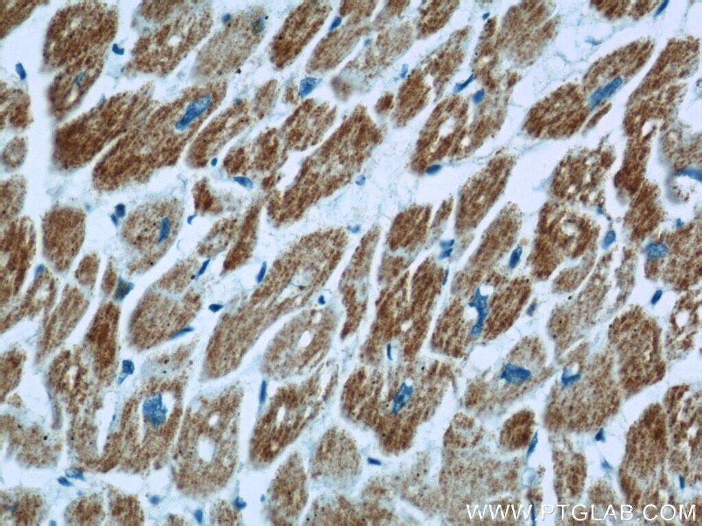
- Immunohistochemistry of paraffin-embedded human heart using 21705-1-AP (UQCRC1 antibody) at dilution of 1:100 (under 10x lens).
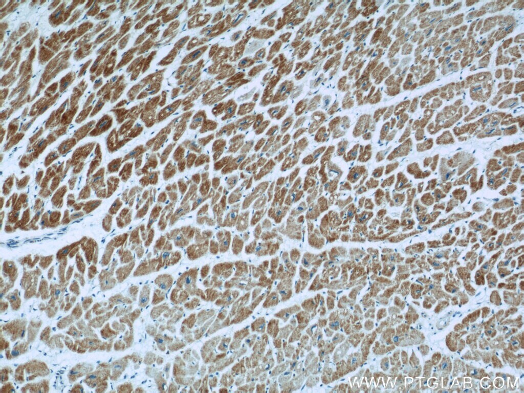
- Submitted by
- Invitrogen Antibodies (provider)
- Main image

- Experimental details
- Immunohistochemistry of paraffin-embedded human heart using 21705-1-AP (UQCRC1 antibody) at dilution of 1:100 (under 40x lens).
Supportive validation
- Submitted by
- Invitrogen Antibodies (provider)
- Main image

- Experimental details
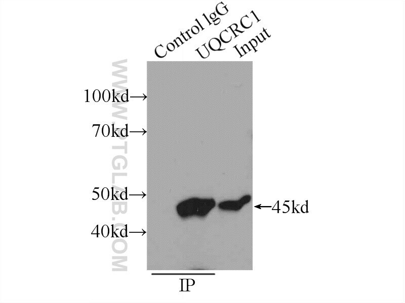
- IP result of anti-UQCRC1 (IP:21705-1-AP, 4ug; Detection:21705-1-AP 1:500) with HeLa cells lysate 1600ug.