Explore
 Validate
 Learn
Learn Western blot
 Immunohistochemistry
Antibody data
- Antibody Data
- Antigen structure
- References [1]
- Comments [0]
- Validations
- Western blot [1]
- Immunohistochemistry [1]
Submit
Validation data
Reference
Comment
Report error
- Product number
- HPA034652 - Provider product page

- Provider
- Atlas Antibodies
- Proper citation
- Atlas Antibodies Cat#HPA034652, RRID:AB_2674263
- Product name
- Anti-PDIA6
- Antibody type
- Polyclonal
- Description
- Polyclonal Antibody against Human PDIA6, Gene description: protein disulfide isomerase family A, member 6, Alternative Gene Names: ERp5, P5, TXNDC7, Validated applications: WB, IHC, Uniprot ID: Q15084, Storage: Store at +4°C for short term storage. Long time storage is recommended at -20°C.
- Reactivity
- Human
- Host
- Rabbit
- Conjugate
- Unconjugated
- Isotype
- IgG
- Vial size
- 100 µl
- Concentration
- 0.2 mg/ml
- Storage
- Store at +4°C for short term storage. Long time storage is recommended at -20°C.
- Handling
- The antibody solution should be gently mixed before use.
Submitted references Protein interaction networks in the vasculature prioritize genes and pathways underlying coronary artery disease
Zhu Q, Hsu Y, Lassen F, MacDonald B, Stead S, Malolepsza E, Kim A, Li T, Mizoguchi T, Schenone M, Guzman G, Tanenbaum B, Fornelos N, Carr S, Gupta R, Ellinor P, Lage K
Communications Biology 2024;7(1)
Communications Biology 2024;7(1)
No comments: Submit comment
Enhanced validation
- Submitted by
- Atlas Antibodies (provider)
- Enhanced method
- Genetic validation
- Main image
- Experimental details
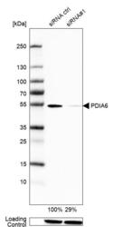
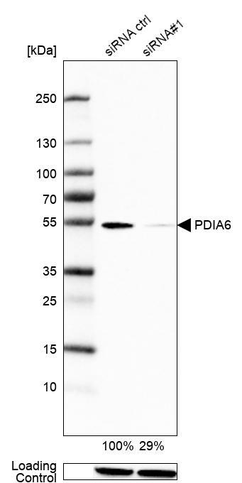
- Western blot analysis in U2OS cells transfected with control siRNA, target specific siRNA probe #1, using Anti-PDIA6 antibody. Remaining relative intensity is presented. Loading control: Anti-GAPDH.
- Sample type
- Human
- Protocol
- Protocol
Supportive validation
- Submitted by
- Atlas Antibodies (provider)
- Enhanced method
- Orthogonal validation
- Main image

- Experimental details
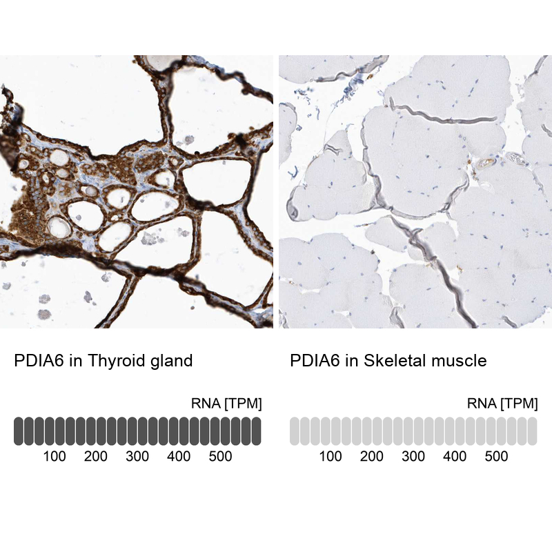
- Immunohistochemistry analysis in human thyroid gland and skeletal muscle tissues using HPA034652 antibody. Corresponding PDIA6 RNA-seq data are presented for the same tissues.
- Sample type
- Human
- Protocol
- Protocol