Explore
 Validate
 Learn
Learn Western blot
 ELISA
ELISAAntibody data
- Antibody Data
- Antigen structure
- References [1]
- Comments [0]
- Validations
- Western blot [1]
Submit
Validation data
Reference
Comment
Report error
- Product number
- A03813-2 - Provider product page

- Provider
- Boster Biological Technology
- Product name
- Anti-PDIA6 Antibody Picoband™
- Antibody type
- Polyclonal
- Description
- Rabbit IgG polyclonal antibody for PDIA6 detection. Tested with WB, IHC-P, ICC/IF, FCM, Direct ELISA in Human;Mouse;Rat.
- Reactivity
- Human, Mouse, Rat
- Host
- Rabbit
- Vial size
- 100μg/vial
- Concentration
- Add 0.2ml of distilled water will yield a concentration of 500ug/ml.
- Storage
- At -20°C for one year. After reconstitution, at 4°C for one month. It can also be aliquotted and stored frozen at -20°C for a longer time. Avoid repeated freezing and thawing.
- Handling
- Add 0.2ml of distilled water will yield a concentration of 500ug/ml.
Submitted references Transcriptomics based identification of S100A3 as the key anti-hepatitis B virus factor of 16F16.
Mahmood F, Xu R, Awan MUN, Jia T, Zhang T, Shi W, Liu M, Han Q, Zhu Q, Zhang Q, Song Y, Xia X, Zhang J
Biomedicine & pharmacotherapy = Biomedecine & pharmacotherapie 2023 Jul;163:114904
Biomedicine & pharmacotherapy = Biomedecine & pharmacotherapie 2023 Jul;163:114904
No comments: Submit comment
Supportive validation
- Submitted by
- Boster Biological Technology (provider)
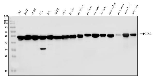
- Main image
- Experimental details
- Western blot analysis of PDIA6 using anti-PDIA6 antibody (A03813-2). Electrophoresis was performed on a 5-20% SDS-PAGE gel at 70V (Stacking gel) / 90V (Resolving gel) for 2-3 hours. The sample well of each lane was loaded with 30ug of sample under reducing conditions. Lane 1: human K562 whole cell lysates, Lane 2: human HepG2 whole cell lysates, Lane 3: human HT1080 whole cell lysates, Lane 4: human Raji whole cell lysates, Lane 5: human Hela whole cell lysates, Lane 6: human HEK293 whole cell lysates, Lane 7: human THP-1 whole cell lysates, Lane 8: human SH-SY5Y whole cell lysates, Lane 9: rat kidney tissue lysates, Lane 10: rat heart tissue lysates, Lane 11: rat liver tissue lysates, Lane 12: rat lung tissue lysates, Lane 13: mouse kidney tissue lysates, Lane 14: mouse heart tissue lysates, Lane 15: mouse liver tissue lysates, Lane 16: mouse lung tissue lysates. After Electrophoresis, proteins were transferred to a Nitrocellulose membrane at 150mA for 50-90 minutes. Blocked the membrane with 5% Non-fat Milk/ TBS for 1.5 hour at RT. The membrane was incubated with rabbit anti-PDIA6 antigen affinity purified polyclonal antibody (Catalog # A03813-2) at 0.25 μg/mL overnight at 4°C, then washed with TBS-0.1%Tween 3 times with 5 minutes each and probed with a goat anti-rabbit IgG-HRP secondary antibody at a dilution of 1:5000 for 1.5 hour at RT. The signal is developed using an Enhanced Chemiluminescent detection (ECL) kit (Catalog # EK1002) with Tanon 5200 system. A specific band was detected for PDIA6 at approximately 50KD. The expected band size for PDIA6 is at 50KD.
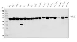
- Additional image