Explore
 Validate
 Learn
Learn Western blot
 Immunocytochemistry
Antibody data
- Antibody Data
- Antigen structure
- References [5]
- Comments [0]
- Validations
- Immunocytochemistry [2]
- Flow cytometry [1]
- Other assay [3]
Submit
Validation data
Reference
Comment
Report error
- Product number
- PA3-008 - Provider product page

- Provider
- Invitrogen Antibodies
- Product name
- PDIA6 Polyclonal Antibody
- Antibody type
- Polyclonal
- Antigen
- Synthetic peptide
- Description
- PA3-008 detects P5 protein in human samples. PA3-008 has successfully been used in Western blot procedures. By Western blot, this antibody detects an ~50 kDa protein representing P5 in human fibrosarcoma HT 1080 cell lysate. The PA3-008 immunogen is a synthetic peptide corresponding to residues D(476) I D L S D V E L D D L G K D E L(492) of human P5. This peptide (Cat. # PEP-224) is available for use in neutralization and control experiments.
- Reactivity
- Human
- Host
- Rabbit
- Isotype
- IgG
- Vial size
- 100 μL
- Concentration
- Conc. Not Determined
- Storage
- -20°C, Avoid Freeze/Thaw Cycles
Submitted references ERp18 regulates activation of ATF6α during unfolded protein response.
Transmembrane thioredoxin-related protein TMX1 is reversibly oxidized in response to protein accumulation in the endoplasmic reticulum.
Cyclophilin C Participates in the US2-Mediated Degradation of Major Histocompatibility Complex Class I Molecules.
Protein disulfide isomerase-P5, down-regulated in the final stage of boar epididymal sperm maturation, catalyzes disulfide formation to inhibit protein function in oxidative refolding of reduced denatured lysozyme.
Multi-chaperone complexes regulate the folding of interferon-gamma in the endoplasmic reticulum.
Oka OB, van Lith M, Rudolf J, Tungkum W, Pringle MA, Bulleid NJ
The EMBO journal 2019 Aug 1;38(15):e100990
The EMBO journal 2019 Aug 1;38(15):e100990
Transmembrane thioredoxin-related protein TMX1 is reversibly oxidized in response to protein accumulation in the endoplasmic reticulum.
Matsuo Y, Hirota K
FEBS open bio 2017 Nov;7(11):1768-1777
FEBS open bio 2017 Nov;7(11):1768-1777
Cyclophilin C Participates in the US2-Mediated Degradation of Major Histocompatibility Complex Class I Molecules.
Chapman DC, Stocki P, Williams DB
PloS one 2015;10(12):e0145458
PloS one 2015;10(12):e0145458
Protein disulfide isomerase-P5, down-regulated in the final stage of boar epididymal sperm maturation, catalyzes disulfide formation to inhibit protein function in oxidative refolding of reduced denatured lysozyme.
Akama K, Horikoshi T, Sugiyama A, Nakahata S, Akitsu A, Niwa N, Intoh A, Kakui Y, Sugaya M, Takei K, Imaizumi N, Sato T, Matsumoto R, Iwahashi H, Kashiwabara S, Baba T, Nakamura M, Toda T
Biochimica et biophysica acta 2010 Jun;1804(6):1272-84
Biochimica et biophysica acta 2010 Jun;1804(6):1272-84
Multi-chaperone complexes regulate the folding of interferon-gamma in the endoplasmic reticulum.
Vandenbroeck K, Martens E, Alloza I
Cytokine 2006 Mar 7;33(5):264-73
Cytokine 2006 Mar 7;33(5):264-73
No comments: Submit comment
Supportive validation
- Submitted by
- Invitrogen Antibodies (provider)
- Main image
- Experimental details
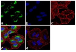
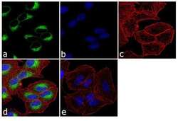
- Immunofluorescence analysis of PDIA6 (P5) was performed using 70% confluent log phase A-549 cells. The cells were fixed with 4% paraformaldehyde for 10 minutes, permeabilized with 0.1% Triton™ X-100 for 10 minutes, and blocked with 1% BSA for 1 hour at room temperature. The cells were labeled with PDIA6 Rabbit Polyclonal Antibody (Product # PA3-008) at 1:250 dilution in 0.1% BSA and incubated for 3 hours at room temperature and then labeled with Goat anti-Rabbit IgG (H+L) Superclonal™ Secondary Antibody, Alexa Fluor® 488 conjugate (Product # A27034) at a dilution of 1:2000 for 45 minutes at room temperature (Panel a: green). Nuclei (Panel b: blue) were stained with SlowFade® Gold Antifade Mountant with DAPI (Product # S36938). F-actin (Panel c: red) was stained with Rhodamine Phalloidin (Product # R415, 1:300). Panel d represents the merged image showing cytoplasmic localization. Panel e shows the no primary antibody control. The images were captured at 60X magnification.
- Submitted by
- Invitrogen Antibodies (provider)
- Main image
- Experimental details
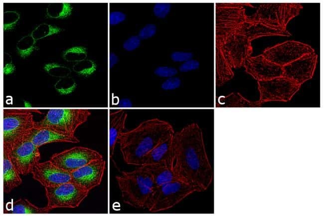
- Immunofluorescence analysis of PDIA6 (P5) was performed using 70% confluent log phase A-549 cells. The cells were fixed with 4% paraformaldehyde for 10 minutes, permeabilized with 0.1% Triton™ X-100 for 10 minutes, and blocked with 1% BSA for 1 hour at room temperature. The cells were labeled with PDIA6 Rabbit Polyclonal Antibody (Product # PA3-008) at 1:250 dilution in 0.1% BSA and incubated for 3 hours at room temperature and then labeled with Goat anti-Rabbit IgG (Heavy Chain) Superclonal™ Secondary Antibody, Alexa Fluor® 488 conjugate (Product # A27034) at a dilution of 1:2000 for 45 minutes at room temperature (Panel a: green). Nuclei (Panel b: blue) were stained with SlowFade® Gold Antifade Mountant with DAPI (Product # S36938). F-actin (Panel c: red) was stained with Rhodamine Phalloidin (Product # R415, 1:300). Panel d represents the merged image showing cytoplasmic localization. Panel e shows the no primary antibody control. The images were captured at 60X magnification.

Supportive validation
- Submitted by
- Invitrogen Antibodies (provider)
- Main image
- Experimental details
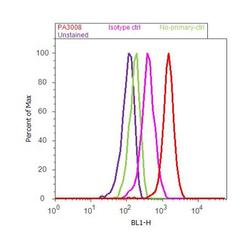
- Flow cytometry analysis of PDIA6 was done on A549 cells. Cells were fixed with 70% ethanol for 10 minutes, permeabilized with 0.25% Tritonª X-100 for 20 minutes, and blocked with 5% BSA for 30 minutes at room temperature. Cells were labeled with PDIA6 Rabbit Polyclonal Antibody (PA3-008, red histogram) or with rabbit isotype control (pink histogram) at 3-5 ug/million cells in 2.5% BSA. After incubation at room temperature for 2 hours, the cells were labeled with Alexa Fluor¨ 488 Goat Anti-Rabbit Secondary Antibody (A11008) at a dilution of 1:400 for 30 minutes at room temperature. The representative 10,000 cells were acquired and analyzed for each sample using an Attune¨ Acoustic Focusing Cytometer. The purple histogram represents unstained control cells and the green histogram represents no-primary-antibody control.

Supportive validation
- Submitted by
- Invitrogen Antibodies (provider)
- Main image
- Experimental details
- NULL

- Submitted by
- Invitrogen Antibodies (provider)
- Main image

- Experimental details
- NULL
- Submitted by
- Invitrogen Antibodies (provider)
- Main image

- Experimental details
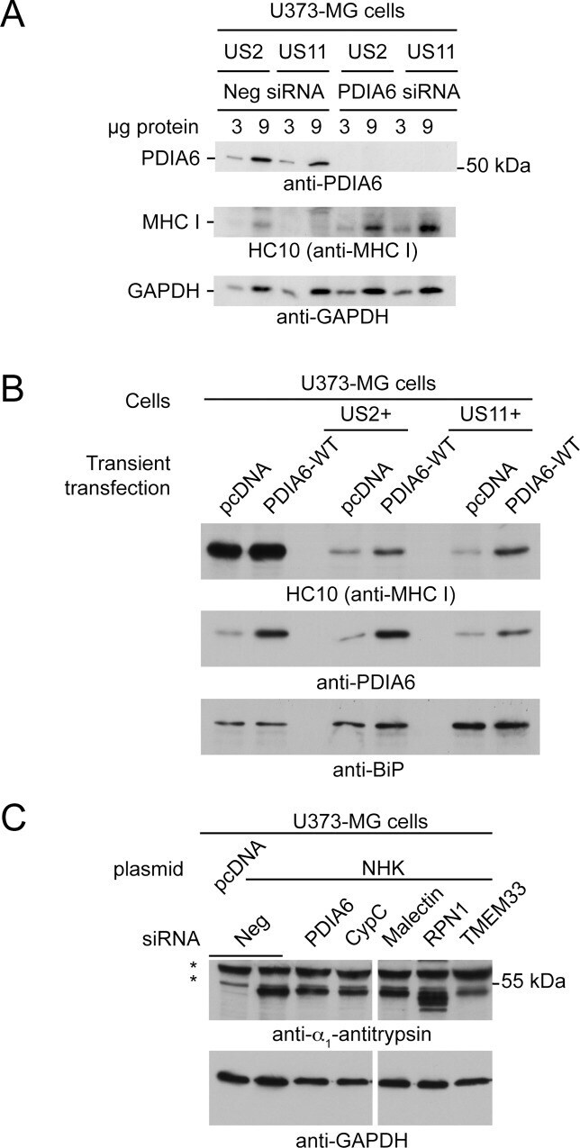
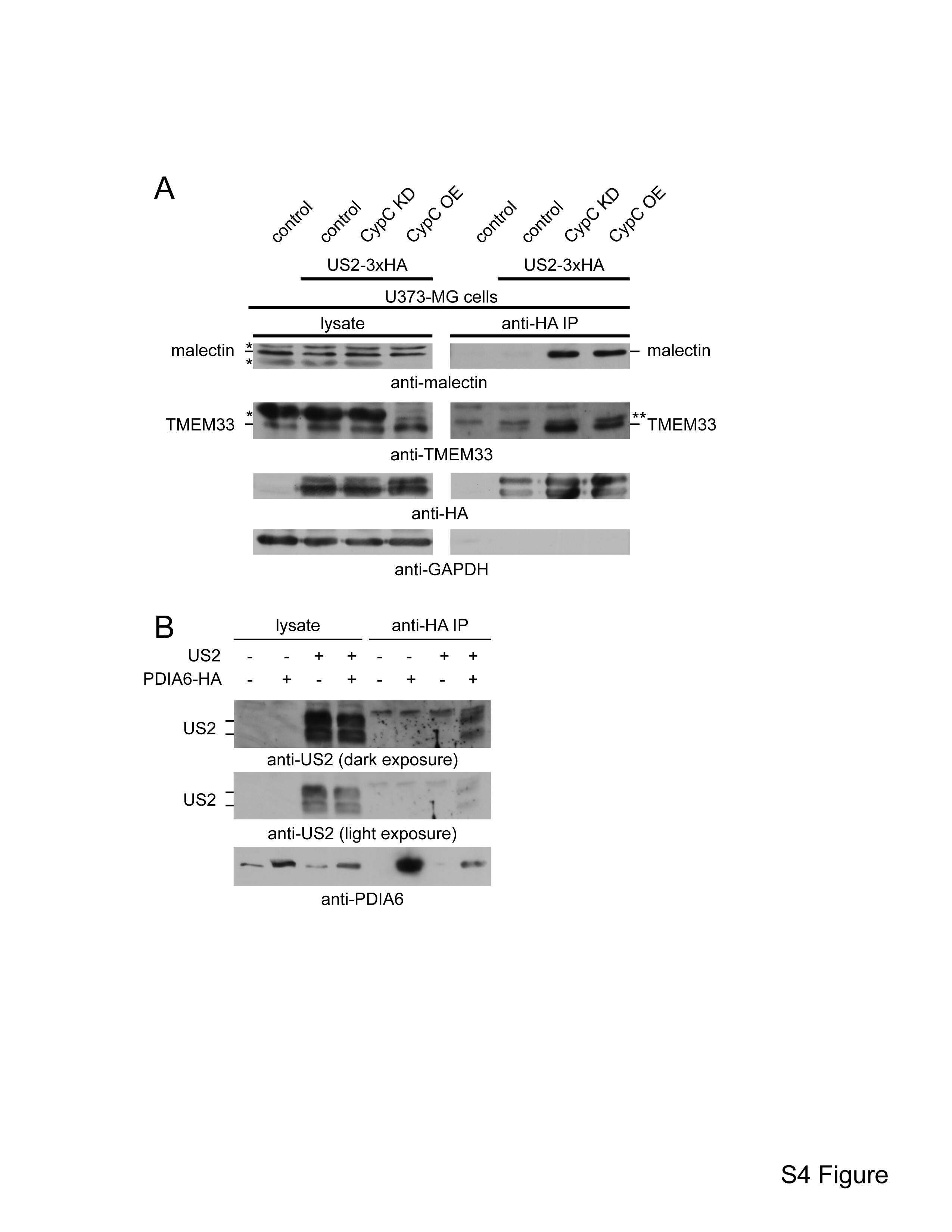
- Fig 7 Involvement of novel US2-associated proteins in other ERAD pathways (A) US11-mediated degradation. U373-MG + US2 or + US11 cells were treated on day 0 and on day 3 with an siRNA specific for PDIA6 or a negative control. On day 6 cells were lysed and MHC I levels were determined by immunoblotting with mAb HC10. GAPDH served as a loading control. (B) U373-MG, U373-MG + US2, or U373-MG + US11 cells were transiently transfected with either empty pcDNA vector or pcDNA + PDIA6 WT . After 48 h, MHC I levels were determined by immunoblot; BiP served as a loading control. (C) U373-MG cells were treated with the indicated panel of pooled siRNAs on days 0 and 3. On day 4 cells were replated and transiently transfected with an alpha 1 -antitrypsin NHK variant expression plasmid. Approximately 48 h later cells were lysed and NHK levels were determined by immunoblotting.