 Explore
 Validate
 Learn
Learn Western blot
 ELISA
ELISAAntibody data
- Antibody Data
- Antigen structure
- References [0]
- Comments [0]
- Validations
- Western blot [5]
- Immunohistochemistry [2]
- Flow cytometry [1]
Submit
Validation data
Reference
Comment
Report error
- Product number
- MA5-17152 - Provider product page

- Provider
- Invitrogen Antibodies
- Product name
- PLK1 Monoclonal Antibody (3C11)
- Antibody type
- Monoclonal
- Antigen
- Purifed from natural sources
- Description
- MA5-17152 targets PLK1 in FACS, IHC, indirect ELISA, and WB applications and shows reactivity with Human and Mouse samples. The MA5-17152 immunogen is purified recombinant fragment of human PLK1 (amino acids: 331-508) expressed in E. Coli. MA5-17152 detects PLK1 which has a predicted molecular weight of approximately 68kDa.
- Reactivity
- Human, Mouse
- Host
- Mouse
- Isotype
- IgG
- Antibody clone number
- 3C11
- Vial size
- 100 µg
- Concentration
- 1 mg/mL
- Storage
- Store at 4°C short term. For long term storage, store at -20°C, avoiding freeze/thaw cycles.
No comments: Submit comment
Supportive validation
- Submitted by
- Invitrogen Antibodies (provider)
- Main image
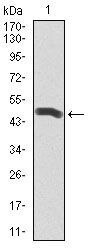
- Experimental details
- Western blot analysis of PLK1 using a PLK1 monoclonal antibody (Product # MA5-17152) against a human PLK1 recombinant protein.

- Submitted by
- Invitrogen Antibodies (provider)
- Main image
- Experimental details
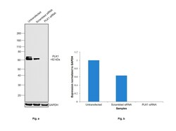
- Knockdown of Serine/threonine-protein kinase PLK1 was achieved by transfecting HeLa with Serine/threonine-protein kinase PLK1 specific siRNAs (Silencer® select Product # s449). Western blot analysis (Fig. a) was performed using Whole cell extracts from the Serine/threonine-protein kinase PLK1 knockdown cells (lane 3), non-targeting scrambled siRNA transfected cells (lane 2) and untransfected cells (lane 1). The blot was probed with PLK1 Monoclonal Antibody (3C11) (Product # MA5-17152, 1:2000 dilution ) and Goat anti-Mouse IgG (H+L) Superclonal™ Recombinant Secondary Antibody, HRP (Product # A28177, 1:4000 dilution). Densitometric analysis of this western blot is shown in histogram (Fig. b). Decrease in signal upon siRNA mediated knockdown confirms that antibody is specific to Serine/threonine-protein kinase PLK1.

- Submitted by
- Invitrogen Antibodies (provider)
- Main image
- Experimental details
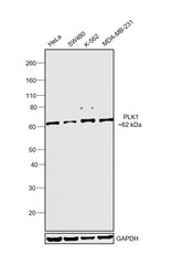
- Western blot was performed using Anti-PLK1 Monoclonal Antibody (3C11) (Product # MA5-17152) and a 62kDa band corresponding to Serine/threonine-protein kinase PLK1 was observed across cell lines tested. Whole cell extracts (30 µg lysate) of HeLa (Lane 1), SW480 (Lane 2), K-562 (Lane 3) and MDA-MB-231 (Lane 4) were electrophoresed using NuPAGE™ 4-12% Bis-Tris Protein Gel (Product # NP0322BOX). Resolved proteins were then transferred onto a Nitrocellulose membrane (Product # IB23001) by iBlot® 2 Dry Blotting System (Product # IB21001). The blot was probed with the primary antibody (1:2000 dilution) and detected by chemiluminescence with Goat anti-Mouse IgG (H+L) Superclonal™ Recombinant Secondary Antibody, HRP (Product # A28177, 1:4000 dilution) using the iBright FL 1000 (Product # A32752). Chemiluminescent detection was performed using SuperSignal™ West Dura Extended Duration Substrate (Product # 34076).

- Submitted by
- Invitrogen Antibodies (provider)
- Main image

- Experimental details
- Western blot analysis of PLK1 using a PLK1 monoclonal antibody (Product # MA5-17152) against a human PLK1 recombinant protein.

- Submitted by
- Invitrogen Antibodies (provider)
- Main image
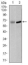
- Experimental details
- Western blot analysis of PLK1 using PLK1 monoclonal antibody (Product # MA5-17152) in K562 (1) and Raji (2) cell lysate.

Supportive validation
- Submitted by
- Invitrogen Antibodies (provider)
- Main image

- Experimental details
- Immunohistochemical analysis of paraffin-embedded stomach cancer tissues using PLK1 monoclonal antibody (Product # MA5-17152) followed with DAB staining.

- Submitted by
- Invitrogen Antibodies (provider)
- Main image

- Experimental details
- Immunohistochemical analysis of paraffin-embedded rectum cancer tissues using PLK1 monoclonal antibody (Product # MA5-17152) followed with DAB staining.

Supportive validation
- Submitted by
- Invitrogen Antibodies (provider)
- Main image

- Experimental details
- Flow cytometric analysis of NIH3T3 cells using PLK1 monoclonal antibody (Product # MA5-17152) (green) and negative control (red).
