Explore
 Validate
 Learn
Learn Western blot
Antibody data
- Antibody Data
- Antigen structure
- References [2]
- Comments [0]
- Validations
- Western blot [2]
- Immunohistochemistry [1]
Submit
Validation data
Reference
Comment
Report error
- Product number
- AM06031PU-T - Provider product page

- Provider
- Acris Antibodies GmbH
- Product name
- anti PLK1 pThr210
- Antibody type
- Monoclonal
- Antigen
- Synthetic peptide, pThr210
- Reactivity
- Human
- Host
- Mouse
- Isotype
- IgG
- Antibody clone number
- 2A3
- Vial size
- 25 µg
- Concentration
- 0.5 mg/ml
Submitted references CDK1 phosphorylation of YAP promotes mitotic defects and cell motility and is essential for neoplastic transformation.
Arpc1b, a centrosomal protein, is both an activator and substrate of Aurora A.
Yang S, Zhang L, Liu M, Chong R, Ding SJ, Chen Y, Dong J
Cancer research 2013 Nov 15;73(22):6722-33
Cancer research 2013 Nov 15;73(22):6722-33
Arpc1b, a centrosomal protein, is both an activator and substrate of Aurora A.
Molli PR, Li DQ, Bagheri-Yarmand R, Pakala SB, Katayama H, Sen S, Iyer J, Chernoff J, Tsai MY, Nair SS, Kumar R
The Journal of cell biology 2010 Jul 12;190(1):101-14
The Journal of cell biology 2010 Jul 12;190(1):101-14
No comments: Submit comment
Supportive validation
- Submitted by
- Acris Antibodies GmbH (provider)
- Main image
- Experimental details
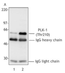
- Panel A: Extracts from untreated Hela cells (Lane 1) or overnight nocodazole-treated Hela cells (Lane 2) were immunoprecipitated with the pan-PLK mAb (clone 3F8), resolved by electrophoresis, transferred to nitrocellulose and probed with mAb 2A3 reactive against Thr210-phosphorylated PLK-1. Proteins were visualized using an HRP goat anti-mouse secondary Ab and a chemiluminescence detection system.

- Submitted by
- Acris Antibodies GmbH (provider)
- Main image

- Experimental details
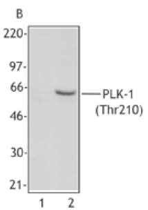
- Panel B: Extracts from untreated Hela cells (Lane 1) or overnight nocodazole-treated Hela cells (Lane 2) were resolved by electrophoresis, transferred to nitrocellulose and probed with mAb 2A3 reactive against Thr210-phosphorylated PLK-1. Proteins were visualized using an HRP goat anti-mouse secondary and a chemiluminescence detection system.
Supportive validation
- Submitted by
- Acris Antibodies GmbH (provider)
- Main image

- Experimental details
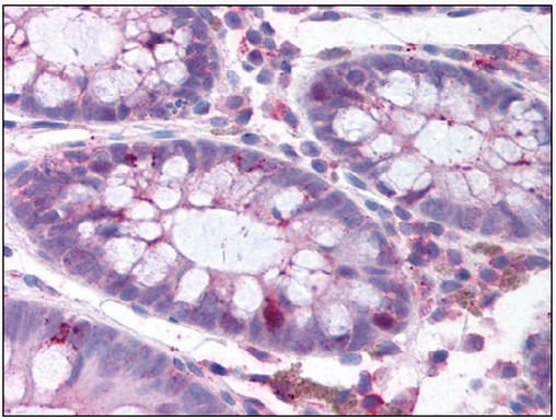
- Formalin-Fixed Paraffin-Embedded Human Colon stained with Phopho-PLK1 Antibody pThr210 Cat.-No AM06031PU (Clone 2A3) at 15 µg/ml after heat-induced antigen retrieval.