 Explore
 Validate
 Learn
Learn Western blot
 Immunoprecipitation
Antibody data
- Antibody Data
- Antigen structure
- References [0]
- Comments [0]
- Validations
- Western blot [3]
- Immunohistochemistry [8]
- Flow cytometry [1]
Submit
Validation data
Reference
Comment
Report error
- Product number
- NBP1-48291 - Provider product page

- Provider
- Novus Biologicals
- Proper citation
- Novus Cat#NBP1-48291, RRID:AB_10010901
- Product name
- Mouse Monoclonal PLK1 Antibody
- Antibody type
- Monoclonal
- Description
- Affinity purified.
- Reactivity
- Human
- Host
- Mouse
- Isotype
- IgG
- Vial size
- 0.1 ml
- Concentration
- 2.5 mg/ml
- Storage
- Store at -20C. Avoid freeze-thaw cycles.
No comments: Submit comment
Supportive validation
- Submitted by
- Novus Biologicals (provider)
- Main image
- Experimental details
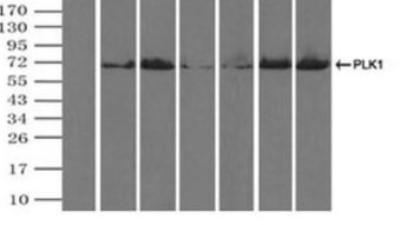
- Western Blot: PLK1 Antibody (1D4) [NBP1-48291] Immunoprecipitation(IP) of PLK1 by using TrueMab monoclonal anti-PLK1 antibodies (Negative control: IP without adding anti-PLK1 antibody.). For each experiment, 500ul of DDK tagged PLK1 overexpression lysates (at 1:5 dilution with HEK293T lysate), 2ug of

- Submitted by
- Novus Biologicals (provider)
- Main image

- Experimental details
- Western Blot: PLK1 Antibody (OTI1D4) [NBP1-48291] - (Negative control: IP without adding anti-PLK1 antibody.). For each experiment, 500ul of DDK tagged PLK1 overexpression lysates (at 1:5 dilution with HEK293T lysate), 2ug of
- Submitted by
- Novus Biologicals (provider)
- Main image

- Experimental details
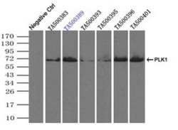
- Western Blot: PLK1 Antibody (OTI1D4) [NBP1-48291] - HEK293T cells were transfected with the pCMV6-ENTRY control (Left lane) or pCMV6-ENTRY PLK1 (Right lane) cDNA for 48 hrs and lysed. Equivalent amounts of cell lysates (5 ug per lane) were separated by SDS-PAGE and immunoblotted with anti-PLK1.
Supportive validation
- Submitted by
- Novus Biologicals (provider)
- Main image

- Experimental details
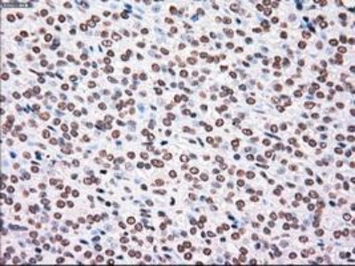
- Immunohistochemistry-Paraffin: PLK1 Antibody (1D4) [NBP1-48291] - Staining of paraffin-embedded Adenocarcinoma of Human endometrium tissue using anti-PLK1 mouse monoclonal antibody.

- Submitted by
- Novus Biologicals (provider)
- Main image

- Experimental details
- Immunohistochemistry-Paraffin: PLK1 Antibody (1D4) [NBP1-48291] - Staining of paraffin-embedded Human endometrium tissue using anti-PLK1 mouse monoclonal antibody.

- Submitted by
- Novus Biologicals (provider)
- Main image

- Experimental details
- Immunohistochemistry-Paraffin: PLK1 Antibody (OTI1D4) [NBP1-48291] - Staining of paraffin-embedded Adenocarcinoma of Human colon tissue using anti-PLK1 mouse monoclonal antibody.

- Submitted by
- Novus Biologicals (provider)
- Main image

- Experimental details
- Immunohistochemistry-Paraffin: PLK1 Antibody (OTI1D4) [NBP1-48291] - Staining of paraffin-embedded Adenocarcinoma of Human endometrium tissue using anti-PLK1 mouse monoclonal antibody.

- Submitted by
- Novus Biologicals (provider)
- Main image

- Experimental details
- Immunohistochemistry-Paraffin: PLK1 Antibody (OTI1D4) [NBP1-48291] - Staining of paraffin-embedded Carcinoma of Human thyroid tissue using anti-PLK1 mouse monoclonal antibody.

- Submitted by
- Novus Biologicals (provider)
- Main image

- Experimental details
- Immunohistochemistry-Paraffin: PLK1 Antibody (OTI1D4) [NBP1-48291] - Staining of paraffin-embedded Human endometrium tissue using anti-PLK1 mouse monoclonal antibody.
- Submitted by
- Novus Biologicals (provider)
- Main image

- Experimental details
- Immunohistochemistry-Paraffin: PLK1 Antibody (OTI1D4) [NBP1-48291] - Staining of paraffin-embedded Human pancreas tissue using anti-PLK1 mouse monoclonal antibody.
- Submitted by
- Novus Biologicals (provider)
- Main image

- Experimental details
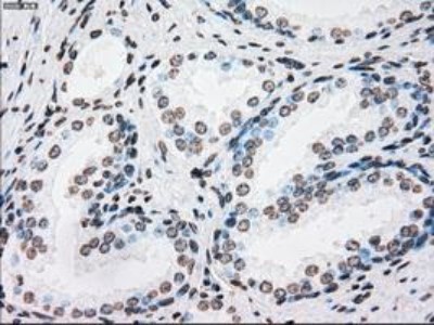
- Immunohistochemistry-Paraffin: PLK1 Antibody (OTI1D4) [NBP1-48291] - Staining of paraffin-embedded Human prostate tissue using anti-PLK1 mouse monoclonal antibody.

Supportive validation
- Submitted by
- Novus Biologicals (provider)
- Main image

- Experimental details
- Flow Cytometry: PLK1 Antibody (OTI1D4) [NBP1-48291] - HEK293T cells transfected with either PLK1 Human Tagged ORF Clone overexpress plasmid (Red), compared to an IgG isotype control, (Green) or empty vector control plasmid (Blue) were immunostained by anti-PLK1 antibody , and then analyzed by flow cytometry (1:100).
