 Explore
 Validate
 Learn
Learn Western blot
 Immunocytochemistry
Antibody data
- Antibody Data
- Antigen structure
- References [15]
- Comments [0]
- Validations
- Immunocytochemistry [5]
- Immunohistochemistry [1]
- Flow cytometry [2]
- Other assay [11]
Submit
Validation data
Reference
Comment
Report error
- Product number
- 459230 - Provider product page

- Provider
- Invitrogen Antibodies
- Product name
- SDHB Monoclonal Antibody (21A11AE7)
- Antibody type
- Monoclonal
- Antigen
- Other
- Description
- Positive control: Human heart mitochondria.
- Reactivity
- Human, Mouse, Rat, Bovine
- Host
- Mouse
- Isotype
- IgG
- Antibody clone number
- 21A11AE7
- Vial size
- 100 μL
- Concentration
- 1 mg/mL
- Storage
- 4°C
Submitted references High-throughput screening for natural compound-based autophagy modulators reveals novel chemotherapeutic mode of action for arzanol.
Herba Houttuyniae Extract Benefits Hyperlipidemic Mice via Activation of the AMPK/PGC-1α/Nrf2 Cascade.
METTL15 introduces N4-methylcytidine into human mitochondrial 12S rRNA and is required for mitoribosome biogenesis.
L-Arabinose Elicits Gut-Derived Hydrogen Production and Ameliorates Metabolic Syndrome in C57BL/6J Mice on High-Fat-Diet.
PIMT/NCOA6IP Deletion in the Mouse Heart Causes Delayed Cardiomyopathy Attributable to Perturbation in Energy Metabolism.
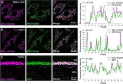
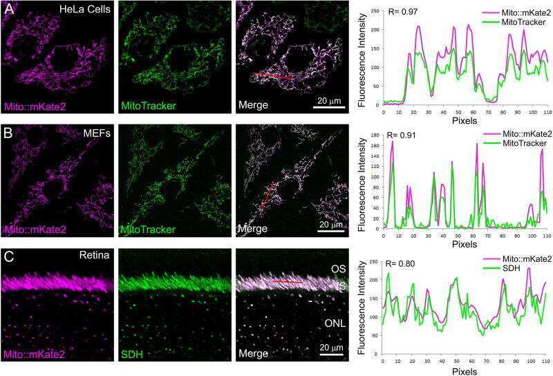
The mito::mKate2 mouse: A far-red fluorescent reporter mouse line for tracking mitochondrial dynamics in vivo.
Mitochondrial Maturation in Human Pluripotent Stem Cell Derived Cardiomyocytes.
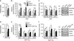
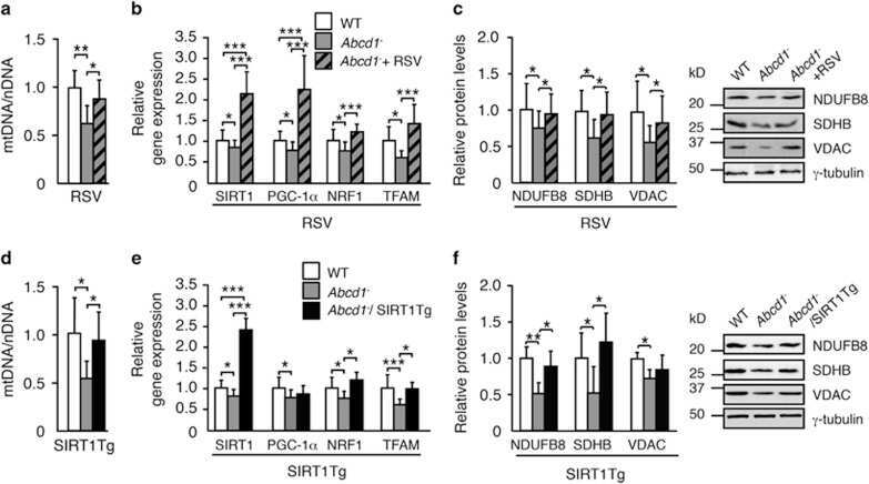
Activation of sirtuin 1 as therapy for the peroxisomal disease adrenoleukodystrophy.
D2HGDH regulates alpha-ketoglutarate levels and dioxygenase function by modulating IDH2.
The transcriptional coregulator PGC-1β controls mitochondrial function and anti-oxidant defence in skeletal muscles.
Prevention and reversal of severe mitochondrial cardiomyopathy by gene therapy in a mouse model of Friedreich's ataxia.
High-throughput screening for growth inhibitors using a yeast model of familial paraganglioma.
Independent roles of methionine sulfoxide reductase A in mitochondrial ATP synthesis and as antioxidant in retinal pigment epithelial cells.
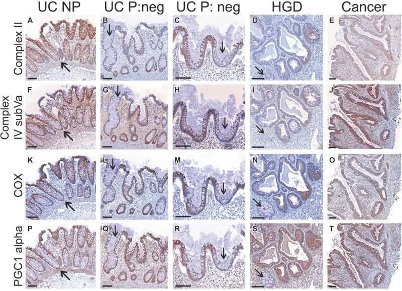
Mitochondria and tumor progression in ulcerative colitis.
β-Adrenergic stimulation does not activate p38 MAP kinase or induce PGC-1α in skeletal muscle.
Deitersen J, Berning L, Stuhldreier F, Ceccacci S, Schlütermann D, Friedrich A, Wu W, Sun Y, Böhler P, Berleth N, Mendiburo MJ, Seggewiß S, Anand R, Reichert AS, Monti MC, Proksch P, Stork B
Cell death & disease 2021 May 31;12(6):560
Cell death & disease 2021 May 31;12(6):560
Herba Houttuyniae Extract Benefits Hyperlipidemic Mice via Activation of the AMPK/PGC-1α/Nrf2 Cascade.
Cao K, Lv W, Liu X, Fan Y, Wang K, Feng Z, Liu J, Zang W, Xing L, Liu J
Nutrients 2020 Jan 7;12(1)
Nutrients 2020 Jan 7;12(1)
METTL15 introduces N4-methylcytidine into human mitochondrial 12S rRNA and is required for mitoribosome biogenesis.
Van Haute L, Hendrick AG, D'Souza AR, Powell CA, Rebelo-Guiomar P, Harbour ME, Ding S, Fearnley IM, Andrews B, Minczuk M
Nucleic acids research 2019 Nov 4;47(19):10267-10281
Nucleic acids research 2019 Nov 4;47(19):10267-10281
L-Arabinose Elicits Gut-Derived Hydrogen Production and Ameliorates Metabolic Syndrome in C57BL/6J Mice on High-Fat-Diet.
Zhao L, Wang Y, Zhang G, Zhang T, Lou J, Liu J
Nutrients 2019 Dec 13;11(12)
Nutrients 2019 Dec 13;11(12)
PIMT/NCOA6IP Deletion in the Mouse Heart Causes Delayed Cardiomyopathy Attributable to Perturbation in Energy Metabolism.
Jia Y, Liu N, Viswakarma N, Sun R, Schipma MJ, Shang M, Thorp EB, Kanwar YS, Thimmapaya B, Reddy JK
International journal of molecular sciences 2018 May 16;19(5)
International journal of molecular sciences 2018 May 16;19(5)
The mito::mKate2 mouse: A far-red fluorescent reporter mouse line for tracking mitochondrial dynamics in vivo.
Barrasso AP, Tong X, Poché RA
Genesis (New York, N.Y. : 2000) 2018 Feb;56(2)
Genesis (New York, N.Y. : 2000) 2018 Feb;56(2)
Mitochondrial Maturation in Human Pluripotent Stem Cell Derived Cardiomyocytes.
Dai DF, Danoviz ME, Wiczer B, Laflamme MA, Tian R
Stem cells international 2017;2017:5153625
Stem cells international 2017;2017:5153625
Activation of sirtuin 1 as therapy for the peroxisomal disease adrenoleukodystrophy.
Morató L, Ruiz M, Boada J, Calingasan NY, Galino J, Guilera C, Jové M, Naudí A, Ferrer I, Pamplona R, Serrano M, Portero-Otín M, Beal MF, Fourcade S, Pujol A
Cell death and differentiation 2015 Nov;22(11):1742-53
Cell death and differentiation 2015 Nov;22(11):1742-53
D2HGDH regulates alpha-ketoglutarate levels and dioxygenase function by modulating IDH2.
Lin AP, Abbas S, Kim SW, Ortega M, Bouamar H, Escobedo Y, Varadarajan P, Qin Y, Sudderth J, Schulz E, Deutsch A, Mohan S, Ulz P, Neumeister P, Rakheja D, Gao X, Hinck A, Weintraub ST, DeBerardinis RJ, Sill H, Dahia PL, Aguiar RC
Nature communications 2015 Jul 16;6:7768
Nature communications 2015 Jul 16;6:7768
The transcriptional coregulator PGC-1β controls mitochondrial function and anti-oxidant defence in skeletal muscles.
Gali Ramamoorthy T, Laverny G, Schlagowski AI, Zoll J, Messaddeq N, Bornert JM, Panza S, Ferry A, Geny B, Metzger D
Nature communications 2015 Dec 17;6:10210
Nature communications 2015 Dec 17;6:10210
Prevention and reversal of severe mitochondrial cardiomyopathy by gene therapy in a mouse model of Friedreich's ataxia.
Perdomini M, Belbellaa B, Monassier L, Reutenauer L, Messaddeq N, Cartier N, Crystal RG, Aubourg P, Puccio H
Nature medicine 2014 May;20(5):542-7
Nature medicine 2014 May;20(5):542-7
High-throughput screening for growth inhibitors using a yeast model of familial paraganglioma.
Bancos I, Bida JP, Tian D, Bundrick M, John K, Holte MN, Her YF, Evans D, Saenz DT, Poeschla EM, Hook D, Georg G, Maher LJ 3rd
PloS one 2013;8(2):e56827
PloS one 2013;8(2):e56827
Independent roles of methionine sulfoxide reductase A in mitochondrial ATP synthesis and as antioxidant in retinal pigment epithelial cells.
Dun Y, Vargas J, Brot N, Finnemann SC
Free radical biology & medicine 2013 Dec;65:1340-1351
Free radical biology & medicine 2013 Dec;65:1340-1351
Mitochondria and tumor progression in ulcerative colitis.
Ussakli CH, Ebaee A, Binkley J, Brentnall TA, Emond MJ, Rabinovitch PS, Risques RA
Journal of the National Cancer Institute 2013 Aug 21;105(16):1239-48
Journal of the National Cancer Institute 2013 Aug 21;105(16):1239-48
β-Adrenergic stimulation does not activate p38 MAP kinase or induce PGC-1α in skeletal muscle.
Kim SH, Asaka M, Higashida K, Takahashi Y, Holloszy JO, Han DH
American journal of physiology. Endocrinology and metabolism 2013 Apr 15;304(8):E844-52
American journal of physiology. Endocrinology and metabolism 2013 Apr 15;304(8):E844-52
No comments: Submit comment
Supportive validation
- Submitted by
- Invitrogen Antibodies (provider)
- Main image
- Experimental details
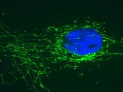
- Immunofluorescent analysis of SDHB using an SDHB Monoclonal antibody (Product # 459230). Cells were fixed, permeabilized, and then labeled with ab14714 followed by an Alexa Fluor® 488-conjugated-goat-anti-mouse IgG2a isotype specific secondary antibody.

- Submitted by
- Invitrogen Antibodies (provider)
- Main image
- Experimental details
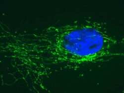
- Immunofluorescent analysis of SDHB using an SDHB Monoclonal antibody (Product # 459230). Cells were fixed, permeabilized, and then labeled with ab14714 followed by an Alexa Fluor® 488-conjugated-goat-anti-mouse IgG2a isotype specific secondary antibody.

- Submitted by
- Invitrogen Antibodies (provider)
- Main image
- Experimental details
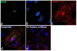
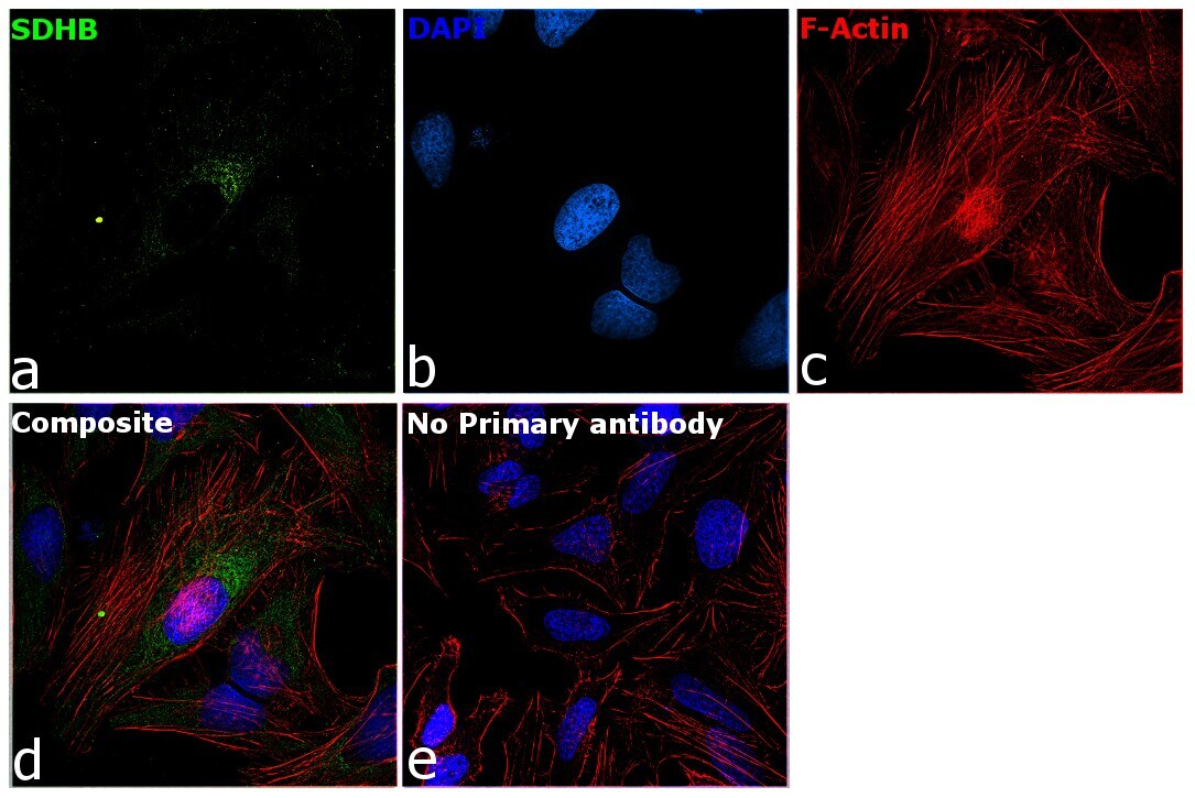
- Immunofluorescence analysis of SDHB was performed using 70% confluent log phase HeLa cells. The cells were fixed with 4% paraformaldehyde for 10 minutes, permeabilized with 0.1% Triton™ X-100 for 15 minutes, and blocked with 2% BSA for 1 hour at room temperature. The cells were labeled with SDHB Monoclonal Antibody (21A11AE7) (Product # 459230) at 5 µg/mL in 0.1% BSA, incubated at 4 degree Celsius overnight and then labeled with Donkey anti-Mouse IgG (H+L) Highly Cross-Adsorbed Secondary Antibody, Alexa Fluor Plus 488 (Product # A32766) at a dilution of 1:2000 for 45 minutes at room temperature (Panel a: green). Nuclei (Panel b: blue) were stained with SlowFade® Gold Antifade Mountant with DAPI (Product # S36938). F-actin (Panel c: red) was stained with Rhodamine Phalloidin (Product # R415, 1:300). Panel d represents the merged image showing mitochondrial localization. Panel e represents control cells with no primary antibody to assess background. The images were captured at 60X magnification.

- Submitted by
- Invitrogen Antibodies (provider)
- Main image

- Experimental details
- Immunofluorescent analysis of SDHB using an SDHB Monoclonal antibody (Product # 459230). Cells were fixed, permeabilized, and then labeled with ab14714 followed by an Alexa Fluor® 488-conjugated-goat-anti-mouse IgG2a isotype specific secondary antibody.

- Submitted by
- Invitrogen Antibodies (provider)
- Main image

- Experimental details
- Immunofluorescence analysis of SDHB was performed using 70% confluent log phase HeLa cells. The cells were fixed with 4% paraformaldehyde for 10 minutes, permeabilized with 0.1% Triton™ X-100 for 15 minutes, and blocked with 2% BSA for 1 hour at room temperature. The cells were labeled with SDHB Monoclonal Antibody (21A11AE7) (Product # 459230) at 5 µg/mL in 0.1% BSA, incubated at 4 degree Celsius overnight and then labeled with Donkey anti-Mouse IgG (H+L) Highly Cross-Adsorbed Secondary Antibody, Alexa Fluor Plus 488 (Product # A32766) at a dilution of 1:2000 for 45 minutes at room temperature (Panel a: green). Nuclei (Panel b: blue) were stained with SlowFade® Gold Antifade Mountant with DAPI (Product # S36938). F-actin (Panel c: red) was stained with Rhodamine Phalloidin (Product # R415, 1:300). Panel d represents the merged image showing mitochondrial localization. Panel e represents control cells with no primary antibody to assess background. The images were captured at 60X magnification.
Supportive validation
- Submitted by
- Invitrogen Antibodies (provider)
- Main image
- Experimental details
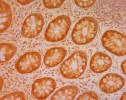
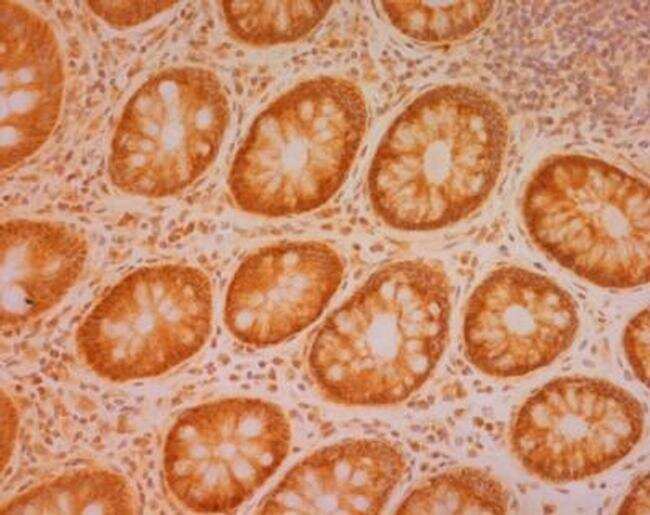
- Immunohistochemical analysis of SDHB in normal aging human colon tissue sections using an SDHB Monoclonal antibody (Product # 459230).
Supportive validation
- Submitted by
- Invitrogen Antibodies (provider)
- Main image

- Experimental details
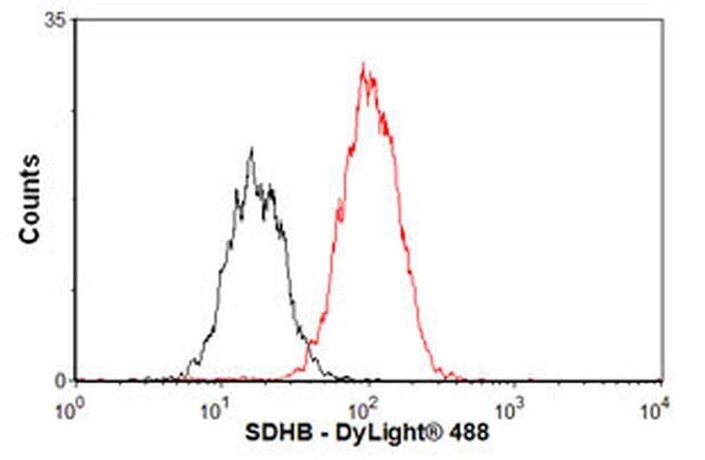
- Flow cytometric analysis of SDHB in HEK293 cells using a SDHB Monoclonal antibody (Product # 459230) at 1 µg/1x10^6 cells, shown in red. The secondary antibody used was DyLight® 488 goat anti-mouse IgG (H+L) at 1:500 dilution. Isotype control, as seen in black, was a mouse IgG2b antibody.
- Submitted by
- Invitrogen Antibodies (provider)
- Main image
- Experimental details
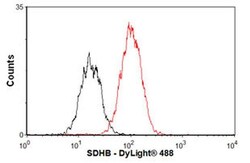
- Flow cytometric analysis of SDHB in HEK293 cells using a SDHB Monoclonal antibody (Product # 459230) at 1 µg/1x10^6 cells, shown in red. The secondary antibody used was DyLight® 488 goat anti-mouse IgG (H+L) at 1:500 dilution. Isotype control, as seen in black, was a mouse IgG2b antibody.

Supportive validation
- Submitted by
- Invitrogen Antibodies (provider)
- Main image
- Experimental details
- NULL
- Submitted by
- Invitrogen Antibodies (provider)
- Main image

- Experimental details
- NULL
- Submitted by
- Invitrogen Antibodies (provider)
- Main image
- Experimental details
- NULL
- Submitted by
- Invitrogen Antibodies (provider)
- Main image
- Experimental details
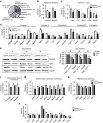
- Figure 5 Impact of myofibre PGC-1beta-deficiency on gene expression. ( a ) Pie chart of gene ontology annotation of genes downregulated by 1.2-fold in gastrocnemius of 8-week-old PGC-1beta (i)skm-/- mice compared with controls. ( b - d ) Relative transcript levels of genes encoding proteins involved in fatty-acid oxidation ( Acadm and Acadvl ; b ) in citric acid cycle ( Dlat , Fh1 , Mdh1 , Pdhb and Idh3b ) ( c ) and Oxidative phosphorylation (OXPHOS; Complex I: Ndufs3 , Ndufv1 , Ndufb3 , Ndufb7 ; Complex II: Sdhb , Sdhd ; Complex III: Cytc , Uqcrc1 , Uqcrfs1 , Uqcr11 ; Complex IV: Cox4i1 , Cox5b ; and Complex V: Atp5i , Atp5o ) ( d ) in gastrocnemius muscle of 8-week-old control and PGC-1beta (i)skm-/- mice, determined by RT-qPCR ( n =8). ( e ) Representative western blot analysis of Ndufs3 (complex I), Sdhb (complex II) and Cytc (complex III) from quadriceps muscle of 8, 18 and 26-week-old control and PGC-1beta (i)skm-/- mice (left panel), and average fold-change (right panel; n =6). Gapdh is used as the internal control. ( f ) Relative transcript levels of Tomm40l , Timm44 and Timm8a1 in gastrocnemius muscle of 8-week-old control and PGC-1beta (i)skm-/- mice, determined by RT-qPCR ( n =8). ( g ) Relative transcript levels of Mtg1 , Tsfm , Gfm1 , Mrpl18 , Mrpl47 , Mrpl55 and Mrps35 in gastrocnemius muscle of 8-week-old control and PGC-1beta (i)skm-/- mice, determined by RT-qPCR ( n =8). ( h ) Relative transcript levels of Mfn1 , Mfn2 , Opa1 , Drp1 and Fis1 in gastrocnemius m
- Submitted by
- Invitrogen Antibodies (provider)
- Main image
- Experimental details
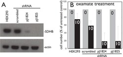
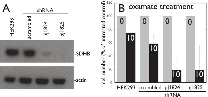
- Figure 11 Mammalian cells knocked down for SDHB expression are differentially sensitive to the lactate dehydrogenase inhibitor oxamate. A. Western blot demonstrating stable lentiviral shRNA knockdown of SDHB in HEK293 cells by SDHB-specific shRNA constructs pJ1824 and pJ1825 but not scrambled shRNA. B. Differential 10 mM oxamate inhibition of HEK293 cell growth after SDHB knockdown.
- Submitted by
- Invitrogen Antibodies (provider)
- Main image
- Experimental details
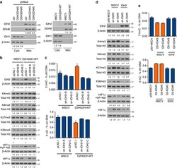
- Figure 6 IDH2 mediates D2HGDH effects on histone and DNA methylation and HIF1alpha hydroxylation. ( a ) Western blot analysis of IDH1 and IDH2 in subcellular fractions of D2HGDH models (knockdown, left panel; ectopic expression, right panel) shows modulation of IDH2 levels. Densitometric quantification is shown at the bottom. ( b ) Top--western blot analysis of IDH2 KD cells expressing an empty-MSCV vector or WT D2HGDH. Middle--Expression of WT D2HGDH decreased H3K methylation (compare lanes marked with a red star); IDH2 KD restored the H3K methylation levels in these cells. Bottom--expression of WT D2HGDH increased HIF1alpha hydroxylation and decreased total HIF1alpha levels (compare lanes marked with a red star); IDH2 KD reversed the increase in HIF1alpha hydroxylation (and decrease in total HIF1alpha) associated with expression of D2HGDH WT. ( c ) Cells expressing D2HGDH-WT (and an empty pLKO.1) displayed a significantly higher abundance of 5hmC marks (top) and a concomitant decrease in global DNA methylation (botton) than MSCV-pLKO.1 controls (marked by red star; P
- Submitted by
- Invitrogen Antibodies (provider)
- Main image
- Experimental details
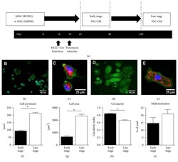
- Figure 1 (a) Differentiation and maturation scheme for early and late stage human ESC-CMs (RuES2) and iPSC-CM (IMR90). hESCs were maintained in a mouse embryonic fibroblast-conditioned medium (MEF-CM) supplemented with fibroblast growth factor (FGF). iPSCs were maintained in mTeSR. Both RuES2 and IMR90 stem cells were differentiated into CMs (day 0) by serial treatment with activin A and BMP-4. On day 14, hESC-CMs were transduced with MCK7-Cre lentivirus during replating onto PEI-gelatin coated glass coverslips/plastic plates. On day 19 cells were replated a second time in the presence of 2 ug/mL puromycin and selected for 48 hours. Cells were maintained for approximately 3 months in culture. We classified cells between days 25 and 40 as early stage PSC-CMs and cells > day 100 as late stage PSC-CMs. Alpha-actinin immunofluorescence (b and d) and mitochondrial staining (red)/eGFP (green) (c and e) of early stage (b-c) and late stage (d-e) hESC-CMs. (f-i) hESC-CM morphometric analysis. * p < 0.05 relative to early stage hESC-CMs, n = 106-121 cells.

- Submitted by
- Invitrogen Antibodies (provider)
- Main image
- Experimental details
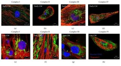
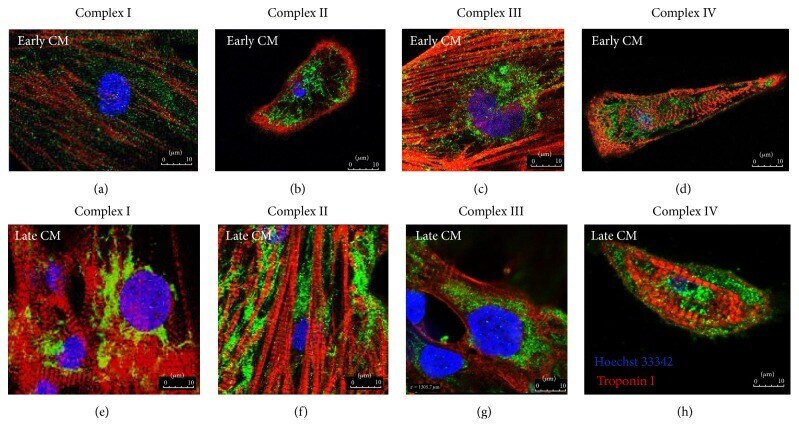
- Figure 4 Immunofluorescence staining of mitochondrial respiratory Complexes I-IV (green), Troponin I (red), and Hoechst 33342 (blue) in early hPSC-CM (a-d) and late hPSC-CM (e-h).
- Submitted by
- Invitrogen Antibodies (provider)
- Main image

- Experimental details
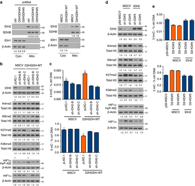
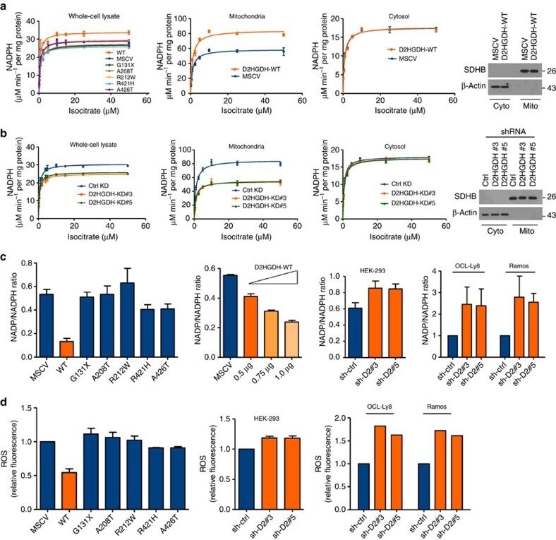
- Figure 5 D2HGDH expression influences mitochondrial IDH activity and the cellular redox state. ( a ) Left panel: HEK-293 cells stably expressing WT D2HGDH displayed a significantly higher V max for IDH than those expressing mutant D2HGDH or an empty vector ( P
- Submitted by
- Invitrogen Antibodies (provider)
- Main image

- Experimental details
- Figure 2. METTL15 is essential for mitochondrial translation. ( A ) Western blot analysis of METTL15 in wild type (WT) and METTL15 KO HAP1 cells. ( B ) Mitochondrial de novo protein synthesis was assessed with 35 S metabolic labelling. Coomassie blue stained (CBS) gel was used as loading control. ( C ) Quantification of the band intensities shown in B using ImageJ. ( D ) Representative example of western blot analysis of NDUFB8, SDHB, UQCRC2, COXII, ATP5A and Beta actin. ( E ) Quantification of 4 western blot experiments for NDUFB8, SDHB, UQCRC2, COXII, ATP5A. Data were statistically analysed by two-tailed Student's t -test. Error bars represent standard deviation of the mean.

- Submitted by
- Invitrogen Antibodies (provider)
- Main image
- Experimental details
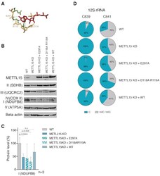
- Figure 5. cDNA complementation of METTL15 KO cells.( A ) Catalytically important residues, D118, R119 and E297, predicted to bind AdoMet (red), that were subjected to site-directed mutagenesis are indicated on the homology model as per Figure 1 . The dotted line represents the predicted interactions with AdoMet based on Wei et al. (2012). ( B ) Western blot analysis of METTL15, NDUFB8 (complex I), SDHB (complex II), UQCRC2 (complex II), COXII (complex IV), ATP5A (complex V) and Beta actin in wild type HAP1 cells, METTL15 KO cells, METTL15 KO cells transfected with cDNA encoding E297A or D118A + R119A mutants or with WT METTL15 cDNA. CBS stands for Coomassie blue staining. ( C ) Quantification of NDUFB8 analysed by western blot ( n = 3, data were statistically analysed by two-tailed Student's t -test. Error bars represent standard deviation of the mean). ( D ) Pie charts of BS RNA-Seq results showing methylation percentage of 12S mt-rRNA position C839 and C841 in wild type HAP1 cells, METTL15 KO cells, and METTL15 KO cells complemented with E297A, D118A + R119A mutants or with WT METTL15 cDNA.
