Explore
 Validate
 Learn
Learn Western blot
 Immunoprecipitation
Antibody data
- Antibody Data
- Antigen structure
- References [0]
- Comments [0]
- Validations
- Western blot [3]
Submit
Validation data
Reference
Comment
Report error
- Product number
- MA5-14964 - Provider product page

- Provider
- Invitrogen Antibodies
- Product name
- TAB1 Monoclonal Antibody (F.211.6)
- Antibody type
- Monoclonal
- Antigen
- Synthetic peptide
- Description
- It is not recommended to aliquot this antibody.
- Reactivity
- Human, Mouse, Rat
- Host
- Rabbit
- Isotype
- IgG
- Antibody clone number
- F.211.6
- Vial size
- 100 µL
- Concentration
- 25 µg/mL
- Storage
- -20°C
No comments: Submit comment
Supportive validation
- Submitted by
- Invitrogen Antibodies (provider)
- Main image
- Experimental details
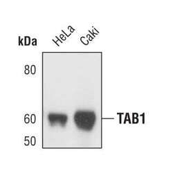
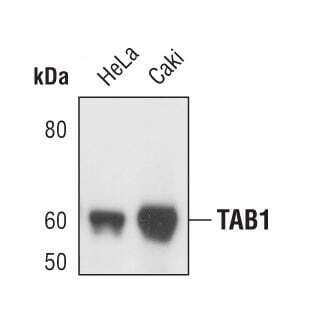
- Western blot analysis of TAB1 in extracts HeLa and Caki cell lines using TAB1 monoclonal antibody (Product # MA5-14964).
- Submitted by
- Invitrogen Antibodies (provider)
- Main image

- Experimental details
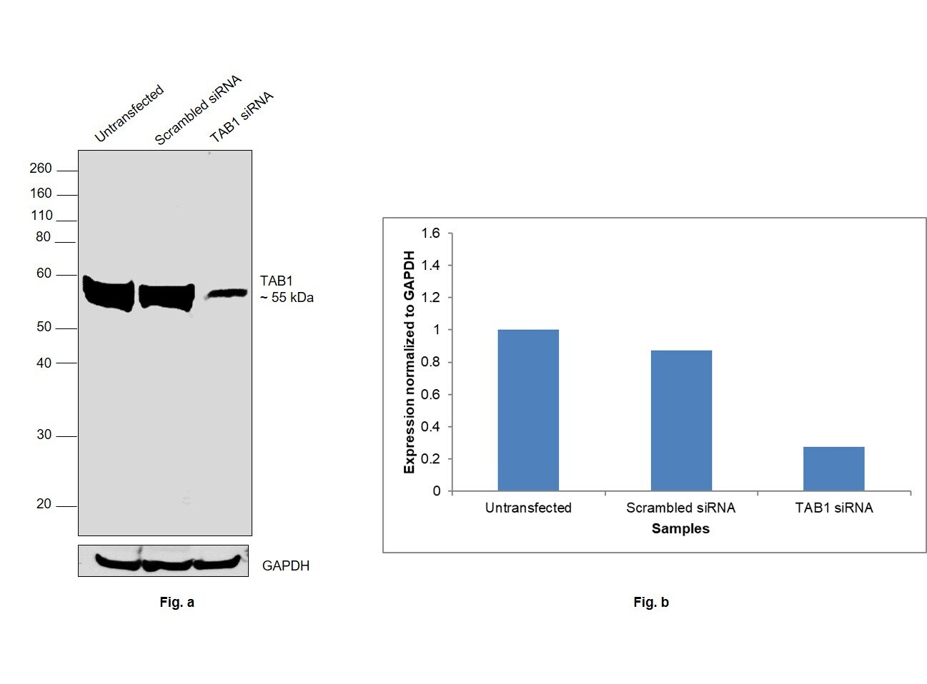
- Knockdown of TAB1 was achieved by transfecting HeLa with TAB1 specific siRNAs (Silencer® select Product # s20451). Western blot analysis (Fig. a) was performed using whole cell extracts from the TAB1 knockdown cells (lane 3), non-specific scrambled siRNA transfected cells (lane 2) and untransfected cells (lane 1). The blot was probed with TAB1 Monoclonal Antibody (Product # MA5-14964, 1:1000 dilution) and Goat anti-Rabbit IgG (H+L), Superclonal™ Recombinant Secondary Antibody, HRP (Product # A27036, 0.25 µg/mL, 1:4000 dilution). Densitometric analysis of this western blot is shown in histogram (Fig. b). Decrease in signal upon siRNA mediated knock down confirms that antibody is specific to TAB1.
- Submitted by
- Invitrogen Antibodies (provider)
- Main image
- Experimental details
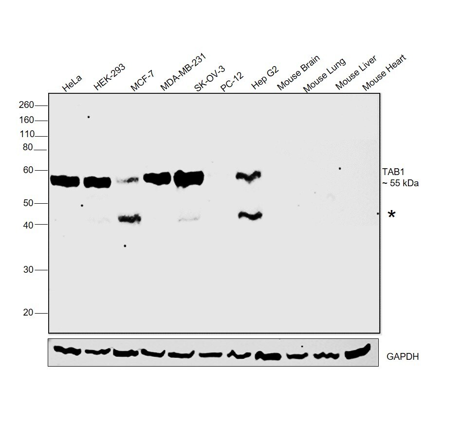
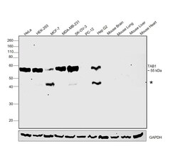
- Western blot was performed using TAB1 Monoclonal Antibody (Product # MA5-14964) and a 55 kDa band corresponding to TAB1 was observed across all the cell lines and tissues tested. An uncharacterized band (*) at~45 kDa was also observed. Whole cell extracts (30 µg lysate) of HeLa (Lane 1), HEK-293 (Lane 2), MCF-7 (Lane 3), MDA-MB-231 (Lane 4), SKOV-3 (Lane 5), PC-12 (Lane 6), HepG2 (Lane 7), tissue extracts of Mouse Brain (Lane 8), Mouse Lung (Lane 9), Mouse Liver (Lane 10) and Mouse Heart (Lane 11) were electrophoresed using NuPAGE™ 10% Bis-Tris Protein Gel (Product # NP0302BOX). Resolved proteins were then transferred onto a nitrocellulose membrane (Product # IB23001) by iBlot® 2 Dry Blotting System (Product # IB21001). The blot was probed with the primary antibody (1:1000 dilution) and detected by chemiluminescence with Goat anti-Rabbit IgG (H+L), Superclonal™ Recombinant Secondary Antibody, HRP (Product # A27036, 1:4000 dilution) using the iBright FL 1000 (Product # A32752). Chemiluminescent detection was performed using Novex® ECL Chemiluminescent Substrate Reagent Kit (Product # WP20005).