Explore
 Validate
 Learn
Learn Western blot
 Immunocytochemistry
Antibody data
- Antibody Data
- Antigen structure
- References [3]
- Comments [0]
- Validations
- Immunocytochemistry [4]
- Immunohistochemistry [1]
- Other assay [1]
Submit
Validation data
Reference
Comment
Report error
- Product number
- PA5-29992 - Provider product page

- Provider
- Invitrogen Antibodies
- Product name
- COX4 Polyclonal Antibody
- Antibody type
- Polyclonal
- Antigen
- Recombinant full-length protein
- Description
- Recommended positive controls: 293T, A431, HeLa, HepG2, mouse muscle, rat muscle, mouse striatal progenitor cells. Predicted reactivity: Rabbit (80%), Rhesus Monkey (93%), Chimpanzee (100%), Bovine (82%). Store product as a concentrated solution. Centrifuge briefly prior to opening the vial.
- Reactivity
- Human, Mouse, Rat
- Host
- Rabbit
- Isotype
- IgG
- Vial size
- 100 μL
- Concentration
- 0.42 mg/mL
- Storage
- Store at 4°C short term. For long term storage, store at -20°C, avoiding freeze/thaw cycles.
Submitted references Nasal Delivery of D-Penicillamine Hydrogel Upregulates a Disintegrin and Metalloprotease 10 Expression via Melatonin Receptor 1 in Alzheimer's Disease Models.
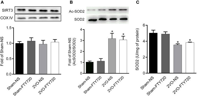
FTY720 Prevents Spatial Memory Impairment in a Rat Model of Chronic Cerebral Hypoperfusion via a SIRT3-Independent Pathway.
Initiation of electron transport chain activity in the embryonic heart coincides with the activation of mitochondrial complex 1 and the formation of supercomplexes.
Zhong M, Kou H, Zhao P, Zheng W, Xu H, Zhang X, Lan W, Guo C, Wang T, Guo F, Wang Z, Gao H
Frontiers in aging neuroscience 2021;13:660249
Frontiers in aging neuroscience 2021;13:660249
FTY720 Prevents Spatial Memory Impairment in a Rat Model of Chronic Cerebral Hypoperfusion via a SIRT3-Independent Pathway.
Zhang M, Hu Y, Zhang J, Zhang J
Frontiers in aging neuroscience 2020;12:593364
Frontiers in aging neuroscience 2020;12:593364
Initiation of electron transport chain activity in the embryonic heart coincides with the activation of mitochondrial complex 1 and the formation of supercomplexes.
Beutner G, Eliseev RA, Porter GA Jr
PloS one 2014;9(11):e113330
PloS one 2014;9(11):e113330
No comments: Submit comment
Supportive validation
- Submitted by
- Invitrogen Antibodies (provider)
- Main image

- Experimental details
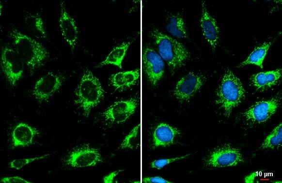
- Immunocytochemistry-Immunofluorescence analysis of COX4 was performed in HeLa cells fixed in ice-cold MeOH for 5 min. Green: COX4 Polyclonal Antibody (Product # PA5-29992) diluted at 1:500. Blue: Hoechst 33342 staining. Scale bar = 10 µm.

- Submitted by
- Invitrogen Antibodies (provider)
- Main image

- Experimental details
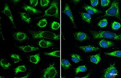
- Immunocytochemistry-Immunofluorescence analysis of COX4 was performed in HeLa cells fixed in ice-cold MeOH for 5 min. Green: COX4 Polyclonal Antibody (Product # PA5-29992) diluted at 1:500. Blue: Hoechst 33342 staining. Scale bar = 10 µm.

- Submitted by
- Invitrogen Antibodies (provider)
- Main image

- Experimental details
- Immunocytochemistry-Immunofluorescence analysis of COX4 was performed in HeLa cells fixed in ice-cold MeOH for 5 min. Green: COX4 Polyclonal Antibody (Product # PA5-29992) diluted at 1:500. Blue: Hoechst 33342 staining. Scale bar = 10 µm.
- Submitted by
- Invitrogen Antibodies (provider)
- Main image
- Experimental details
- Immunocytochemistry-Immunofluorescence analysis of COX4 was performed in HeLa cells fixed in ice-cold MeOH for 5 min. Green: COX4 Polyclonal Antibody (Product # PA5-29992) diluted at 1:500. Blue: Hoechst 33342 staining. Scale bar = 10 µm.

Supportive validation
- Submitted by
- Invitrogen Antibodies (provider)
- Main image
- Experimental details
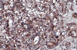
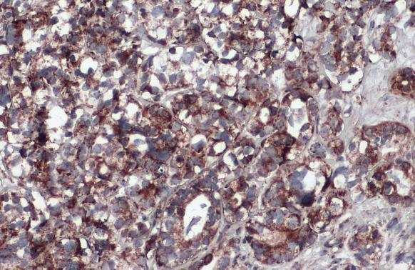
- COX4 Polyclonal Antibody detects COX4 protein at mitochondria by immunohistochemical analysis. Sample: Paraffin-embedded human endometrial carcinoma. COX4 stained by COX4 Polyclonal Antibody (Product # PA5-29992) diluted at 1:500. Antigen Retrieval: Citrate buffer, pH 6.0, 15 min.
Supportive validation
- Submitted by
- Invitrogen Antibodies (provider)
- Main image
- Experimental details
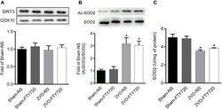
- Figure 6 Representative SIRT3 (A) , acetylated SOD2 K68 (Ac-SOD2 K68) (B) expressions, SOD2 activity (C) in the hippocampus of rats in each group. SIRT3 and SOD2 K68 expression in the hippocampus were normalized to loading of COX IV and SOD2, respectively, and were expressed as fold of sham-NS. Data were shown as mean +- S.E.M. n = 4-5 rats for each group. * p < 0.05 compared with the sham-NS group.