Explore
 Validate
 Learn
Learn Western blot
Antibody data
- Antibody Data
- Antigen structure
- References [4]
- Comments [0]
- Validations
- Western blot [3]
- Other assay [6]
Submit
Validation data
Reference
Comment
Report error
- Product number
- PA5-19521 - Provider product page

- Provider
- Invitrogen Antibodies
- Product name
- ATF4 Polyclonal Antibody
- Antibody type
- Polyclonal
- Antigen
- Synthetic peptide
- Description
- Application Note: For IHC, epitope retrieval with citrate buffer pH 6.0 is recommended for FFPE tissue sections.
- Reactivity
- Human, Mouse, Rat
- Host
- Rabbit
- Isotype
- IgG
- Vial size
- 100 µg
- Concentration
- 0.8 mg/mL
- Storage
- Store at 4°C short term. For long term storage, store at -20°C, avoiding freeze/thaw cycles.
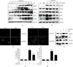
Submitted references Anthocyanins from Hibiscus syriacus L. Inhibit NLRP3 Inflammasome in BV2 Microglia Cells by Alleviating NF-κB- and ER Stress-Induced Ca(2+) Accumulation and Mitochondrial ROS Production.
Fisetin Protects HaCaT Human Keratinocytes from Fine Particulate Matter (PM(2.5))-Induced Oxidative Stress and Apoptosis by Inhibiting the Endoplasmic Reticulum Stress Response.
Protective Effect of Anthocyanin-Enriched Polyphenols from Hibiscus syriacus L. (Malvaceae) against Ultraviolet B-Induced Damage.
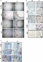
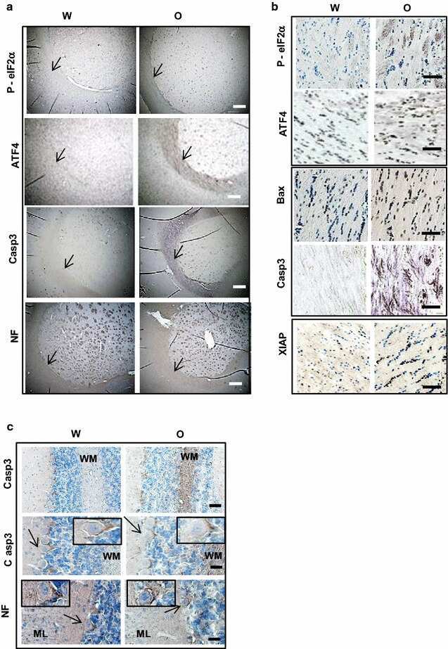
Chronic oxycodone induces axonal degeneration in rat brain.
Molagoda IMN, Lee KT, Choi YH, Jayasingha JACC, Kim GY
Oxidative medicine and cellular longevity 2021;2021:1246491
Oxidative medicine and cellular longevity 2021;2021:1246491
Fisetin Protects HaCaT Human Keratinocytes from Fine Particulate Matter (PM(2.5))-Induced Oxidative Stress and Apoptosis by Inhibiting the Endoplasmic Reticulum Stress Response.
Molagoda IMN, Kavinda MHD, Choi YH, Lee H, Kang CH, Lee MH, Lee CM, Kim GY
Antioxidants (Basel, Switzerland) 2021 Sep 18;10(9)
Antioxidants (Basel, Switzerland) 2021 Sep 18;10(9)
Protective Effect of Anthocyanin-Enriched Polyphenols from Hibiscus syriacus L. (Malvaceae) against Ultraviolet B-Induced Damage.
Karunarathne WAHM, Molagoda IMN, Lee KT, Choi YH, Yu SM, Kang CH, Kim GY
Antioxidants (Basel, Switzerland) 2021 Apr 9;10(4)
Antioxidants (Basel, Switzerland) 2021 Apr 9;10(4)
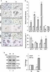
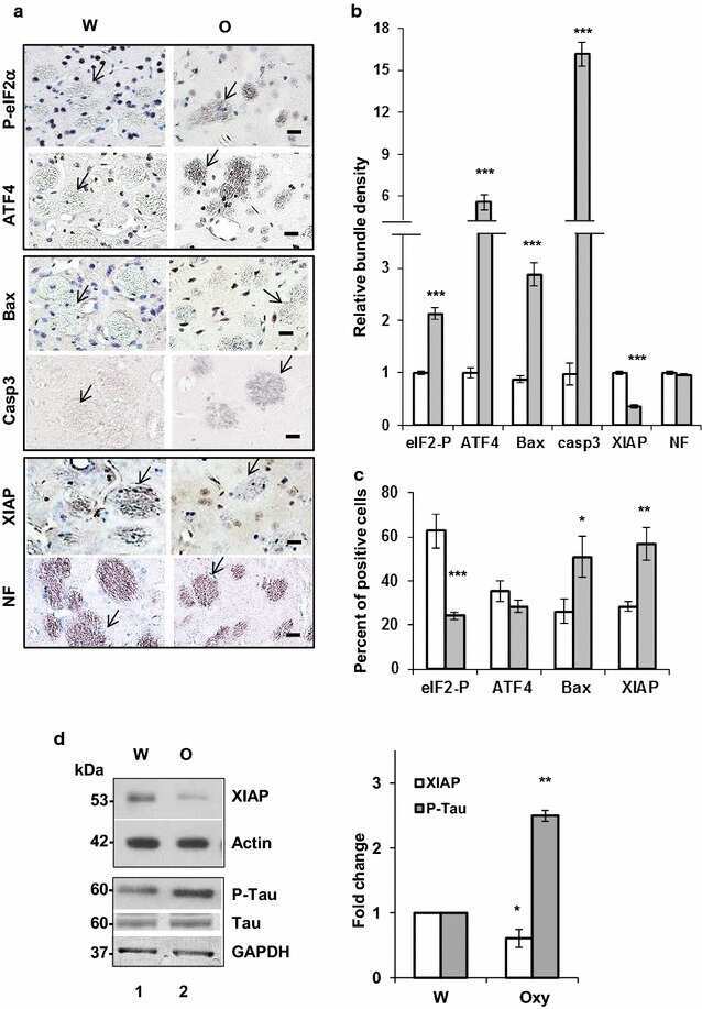
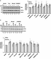
Chronic oxycodone induces axonal degeneration in rat brain.
Fan R, Schrott LM, Arnold T, Snelling S, Rao M, Graham D, Cornelius A, Korneeva NL
BMC neuroscience 2018 Mar 23;19(1):15
BMC neuroscience 2018 Mar 23;19(1):15
No comments: Submit comment
Supportive validation
- Submitted by
- Invitrogen Antibodies (provider)
- Main image
- Experimental details
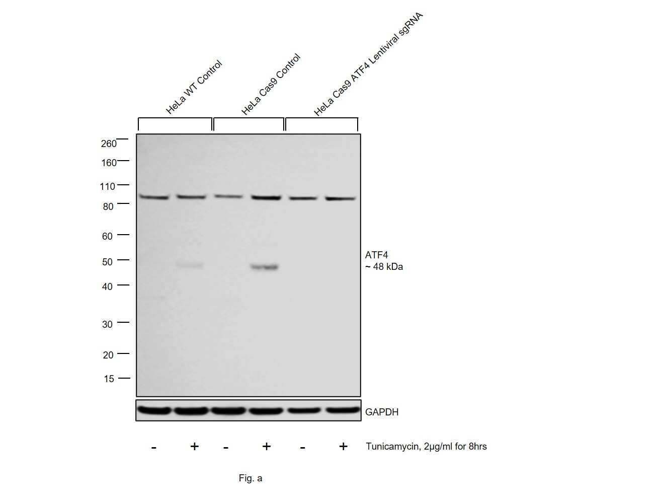
- CRISPR-Cas9 mediated genome editing of ATF4 (as confirmed by next generation sequencing) was achieved by using LentiArray™ Lentiviral sgRNA (Product # A32042, Assay ID CRISPR988840_LV) and LentiArray Cas9 Lentivirus (Product # A32064). Fig (a) Western blot analysis of ATF4 was performed by loading 30 µg of HeLa Wild Type (Lane 1), HeLa Wild type treated with 2 µg/mL tunicamycin for 8 hrs (Lane 2), HeLa Cas9 (Lane 3), HeLa Cas9 treated with 2 µg/mL tunicamycin for 8 hrs (Lane 4), HeLa Cas9 transduced with ATF4 Lentiviral sgRNA (Lane 5) and HeLa Cas9 transduced with ATF4 Lentiviral sgRNA and treated with 2 µg/mL tunicamycin for 8 hrs cells (Lane 6) whole cell extracts. The samples were electrophoresed using NuPAGE™ Novex™ 4-12% Bis-Tris Protein Gel (Product # NP0322BOX). Resolved proteins were then transferred onto a nitrocellulose membrane (Product # IB23001) by iBlot® 2 Dry Blotting System (Product # IB21001). The blot was probed with an ATF4 Polyclonal Antibody (Product # PA5-19521) using 1 µg/mL dilution and Goat anti-Rabbit IgG (H+L) Superclonal™ Recombinant Secondary Antibody, HRP (Product # A27036 1:8,000 dilution). Chemiluminescent detection was performed using SuperSignal™ West Dura Extended Duration Substrate (Product # 34076). A loss of signal in sgRNA transduced cells using the LentiArray™ CRISPR product line confirms that antibody is specific to ATF4. An uncharacterized band at ~90 kDa was obser
- Submitted by
- Invitrogen Antibodies (provider)
- Main image
- Experimental details
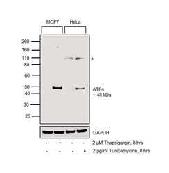
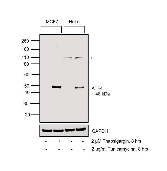
- Western blot was performed using Anti-ATF4 Polyclonal Antibody (Product # PA5-19521) and a 48 kDa band corresponding to ATF4 was observed across cell lines tested. Whole cell extracts (30 µg lysate) of MCF7 (Lane 1), MCF7 treated with 2 µM Thapsigargin for 8 hours (Lane 2), HeLa (Lane 3) and HeLa treated with 2 µg/ml Tunicamycin for 8 hours (Lane 4) were electrophoresed using NuPAGE™ 4-12% Bis-Tris Protein Gel (Product # NP0322BOX). Resolved proteins were then transferred onto a Nitrocellulose membrane (Product # IB23001) by iBlot® 2 Dry Blotting System (Product # IB21001). The blot was probed with the primary antibody (1 µg/mL) and detected by chemiluminescence with Goat anti-Rabbit IgG (H+L) Superclonal™ Recombinant Secondary Antibody, HRP (Product # A27036,1:4000) using the iBright FL 1000 (Product # A32752). Chemiluminescent detection was performed using Novex® ECL Chemiluminescent Substrate Reagent Kit (Product # WP20005). An uncharacterized band (*) at 110 kDa was observed.
- Submitted by
- Invitrogen Antibodies (provider)
- Main image
- Experimental details
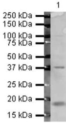
- Western blot analysis of HeLa Whole Cell Lysate using Product # PA5-19521, ATF4 primary antibody at a dilution of 1 µg/mL. Blot treated with a secondary IR Dye680-conjugated Goat polyclonal anti-Rabbit antibody was used at a dilution of 1:10000.

Supportive validation
- Submitted by
- Invitrogen Antibodies (provider)
- Main image
- Experimental details
- NULL
- Submitted by
- Invitrogen Antibodies (provider)
- Main image
- Experimental details
- NULL
- Submitted by
- Invitrogen Antibodies (provider)
- Main image
- Experimental details
- NULL

- Submitted by
- Invitrogen Antibodies (provider)
- Main image
- Experimental details
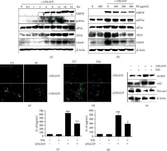
- Figure 3 PS inhibit the LPS/ATP-induced NLRP3 inflammasome by downregulating ER stress-induced Ca 2+ accumulation. (a) BV2 microglia cells were stimulated with LPS/ATP. Total proteins were isolated, and western blotting was performed. (b) In a parallel experiment, the cells were pretreated with the indicated concentrations of PS (0-400 mu g/mL) for 2 h prior to stimulation with LPS/ATP for 12 h. Total protein was extracted, and western blotting was performed. BV2 microglia cells were pretreated with (c) PS (400 mu g/mL) and (d) salubrinal (SAL, 10 mu M) for 2 h prior to stimulation with LPS/ATP for 12 h. The cells were stained with 1 mu M Fluo-4 AM, and cell images were captured using CELENA S Digital Imaging System. (e) Under SAL-treated conditions, the total protein was isolated at 12 h, and western blotting was performed. Cell culture supernatants were collected 48 h after treatment with LPS/ATP, and ELISA was performed to quantify the levels of (f) IL-1 beta and (g) IL-18. ### p < 0.001 vs. untreated cells; *** p < 0.001 and * p < 0.05 vs. LPS/ATP-treated cells. UT: untreated cells.
- Submitted by
- Invitrogen Antibodies (provider)
- Main image

- Experimental details
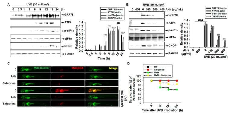
- Figure 8 Effect of anthocyanins from the flower petals of Hibiscus syriacus L. (Malvaceae, AHs) on ER stress and mtROS production. ( A ) and ( B ) The expression of GRP78, ATF4, p-eIF1alpha, CHOP, and beta-actin protein (left) and relative density (right). ( C ) The staining of MitoSOX Red and MitoTracker Green. ( D ) Survival rate of zebrafish. * p < 0.05 and *** p < 0.001 vs. UVB-irradiated cells and ### p < 0.001 vs. untreated cells (UT).
- Submitted by
- Invitrogen Antibodies (provider)
- Main image

- Experimental details
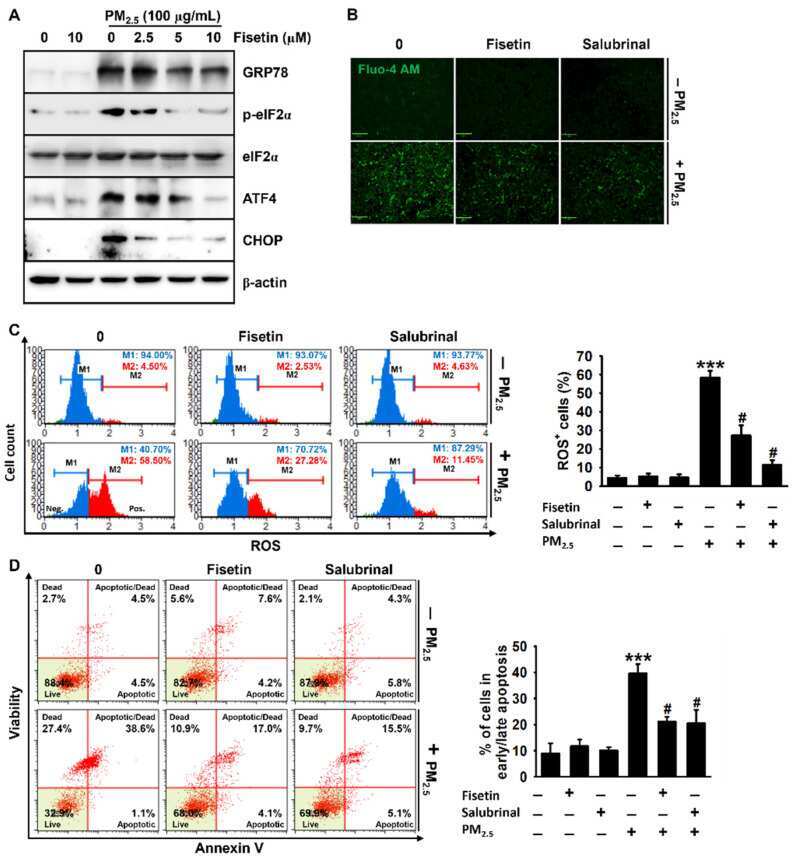
- Figure 4 Fisetin inhibits PM 2.5 -induced apoptosis by alleviating ER stress. HaCaT keratinocytes were treated with fisetin (0-10 uM) for 2 h and subsequently exposed to 100 ug/mL PM 2.5 for 24 h. ( A ) The total proteins were extracted, and Western blotting was performed for detecting the expression of GRP78, p-eIF2alpha, eIF2alpha, ATF4, and CHOP. beta-Actin was used as the loading control. ( B ) The cells were treated with 10 uM fisetin or 20 uM salubrinal in the presence or absence of 100 ug/mL PM 2.5 for 24 h. The cells were incubated with Ca 2+ -sensitive Fluo-4 AM for 10 min and live images were captured using a CELENA S Digital Imaging System. Scale bar = 100 um. ( C , D ) In a parallel experiment, the cells were treated with 10 uM fisetin or 20 uM salubrinal in the presence or absence of 100 ug/mL PM 2.5 for 24 h. The cells were stained with a ( C ) Muse Oxidative Stress Assay Kit and ( D ) Muse Annexin V & Dead Cell Assay Kit. *** p < 0.001 vs. untreated cells and # p < 0.05 vs. PM 2.5 -treated cells.