Explore
 Validate
 Learn
Learn Western blot
 Immunocytochemistry
Antibody data
- Antibody Data
- Antigen structure
- References [4]
- Comments [0]
- Validations
- Immunocytochemistry [5]
- Immunohistochemistry [1]
- Other assay [4]
Submit
Validation data
Reference
Comment
Report error
- Product number
- PA5-21677 - Provider product page

- Provider
- Invitrogen Antibodies
- Product name
- NDUFS4 Polyclonal Antibody
- Antibody type
- Polyclonal
- Antigen
- Recombinant full-length protein
- Description
- Recommended positive controls: 293T, mouse brain, rat heart. Predicted reactivity: Mouse (86%), Rat (86%), Zebrafish (80%), Chimpanzee (99%), Bovine (89%). Store product as a concentrated solution. Centrifuge briefly prior to opening the vial.
- Reactivity
- Human, Mouse, Rat
- Host
- Rabbit
- Isotype
- IgG
- Vial size
- 100 μL
- Concentration
- 1 mg/mL
- Storage
- Store at 4°C short term. For long term storage, store at -20°C, avoiding freeze/thaw cycles.
Submitted references Mitochondrial calcium uniporter stabilization preserves energetic homeostasis during Complex I impairment.
cAMP/PKA Signaling Modulates Mitochondrial Supercomplex Organization.
Enhancing natriuretic peptide signaling in adipose tissue, but not in muscle, protects against diet-induced obesity and insulin resistance.
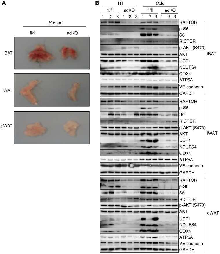
Activation of mTORC1 is essential for β-adrenergic stimulation of adipose browning.
Balderas E, Eberhardt DR, Lee S, Pleinis JM, Sommakia S, Balynas AM, Yin X, Parker MC, Maguire CT, Cho S, Szulik MW, Bakhtina A, Bia RD, Friederich MW, Locke TM, Van Hove JLK, Drakos SG, Sancak Y, Tristani-Firouzi M, Franklin S, Rodan AR, Chaudhuri D
Nature communications 2022 May 19;13(1):2769
Nature communications 2022 May 19;13(1):2769
cAMP/PKA Signaling Modulates Mitochondrial Supercomplex Organization.
Signorile A, Pacelli C, Palese LL, Santeramo A, Roca E, Cocco T, De Rasmo D
International journal of molecular sciences 2022 Aug 25;23(17)
International journal of molecular sciences 2022 Aug 25;23(17)
Enhancing natriuretic peptide signaling in adipose tissue, but not in muscle, protects against diet-induced obesity and insulin resistance.
Wu W, Shi F, Liu D, Ceddia RP, Gaffin R, Wei W, Fang H, Lewandowski ED, Collins S
Science signaling 2017 Jul 25;10(489)
Science signaling 2017 Jul 25;10(489)
Activation of mTORC1 is essential for β-adrenergic stimulation of adipose browning.
Liu D, Bordicchia M, Zhang C, Fang H, Wei W, Li JL, Guilherme A, Guntur K, Czech MP, Collins S
The Journal of clinical investigation 2016 May 2;126(5):1704-16
The Journal of clinical investigation 2016 May 2;126(5):1704-16
No comments: Submit comment
Supportive validation
- Submitted by
- Invitrogen Antibodies (provider)
- Main image
- Experimental details
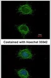
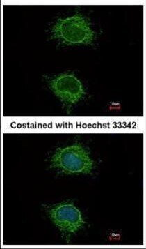
- Immunofluorescent analysis of NDUFS4 in methanol-fixed HeLa cells using a NDUFS4 polyclonal antibody (Product # PA5-21677) at a 1:100 dilution.
- Submitted by
- Invitrogen Antibodies (provider)
- Main image
- Experimental details
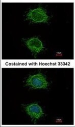
- Immunofluorescence analysis of methanol-fixed HeLa, using NDUFS4 antibody (Product # PA5-21677) at 1:100 dilution.
- Submitted by
- Invitrogen Antibodies (provider)
- Main image
- Experimental details
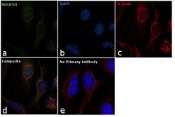
- Immunofluorescence analysis of NDUFS4 was performed using 70% confluent log phase HeLa cells. The cells were fixed with 4% paraformaldehyde for 10 minutes, permeabilized with 0.1% Triton™ X-100 for 10 minutes, and blocked with 1% BSA for 1 hour at room temperature. The cells were labeled with NDUFS4 Rabbit Polyclonal Antibody (Product # PA5-21677) at 5 µg/mL in 0.1% BSA and incubated overnight at 4 degree and then labeled with Goat anti-Rabbit IgG (H+L) Superclonal™ Secondary Antibody, Alexa Fluor® 488 conjugate (Product # A27034) at a dilution of 1:2000 for 45 minutes at room temperature (Panel a: green). Nuclei (Panel b: blue) were stained with SlowFade® Gold Antifade Mountant with DAPI (Product # S36938). Mitochondria (Panel c: red) was stained with Mitotracker Red CMXRos (Product # M7512). Panel d represents the merged image showing Mitochondrial localization. Panel e represents control cells with no primary antibody to assess background. The images were captured at 60X magnification.
- Submitted by
- Invitrogen Antibodies (provider)
- Main image

- Experimental details
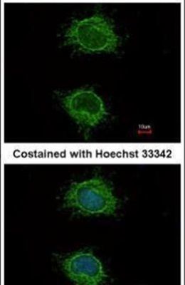
- Immunofluorescence analysis of methanol-fixed HeLa, using NDUFS4 antibody (Product # PA5-21677) at 1:100 dilution.

- Submitted by
- Invitrogen Antibodies (provider)
- Main image
- Experimental details
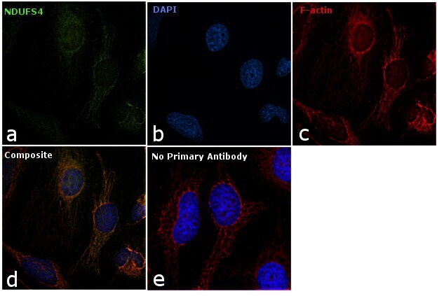
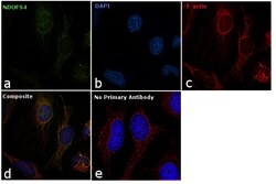
- Immunofluorescence analysis of NDUFS4 was performed using 70% confluent log phase HeLa cells. The cells were fixed with 4% paraformaldehyde for 10 minutes, permeabilized with 0.1% Triton™ X-100 for 10 minutes, and blocked with 1% BSA for 1 hour at room temperature. The cells were labeled with NDUFS4 Rabbit Polyclonal Antibody (Product # PA5-21677) at 5 µg/mL in 0.1% BSA and incubated overnight at 4 degree and then labeled with Goat anti-Rabbit IgG (Heavy Chain) Superclonal™ Secondary Antibody, Alexa Fluor® 488 conjugate (Product # A27034) at a dilution of 1:2000 for 45 minutes at room temperature (Panel a: green). Nuclei (Panel b: blue) were stained with SlowFade® Gold Antifade Mountant with DAPI (Product # S36938). Mitochondria (Panel c: red) was stained with Mitotracker Red CMXRos (Product # M7512). Panel d represents the merged image showing Mitochondrial localization. Panel e represents control cells with no primary antibody to assess background. The images were captured at 60X magnification.

Supportive validation
- Submitted by
- Invitrogen Antibodies (provider)
- Main image

- Experimental details
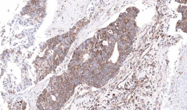
- Immunohistochemical analysis of paraffin-embedded human ovarian cancer, using NDUFS4 (Product # PA5-21677) antibody at 1:100 dilution. Antigen Retrieval: EDTA based buffer, pH 8.0, 15 min.
Supportive validation
- Submitted by
- Invitrogen Antibodies (provider)
- Main image
- Experimental details
- NULL
- Submitted by
- Invitrogen Antibodies (provider)
- Main image
- Experimental details
- NULL
- Submitted by
- Invitrogen Antibodies (provider)
- Main image
- Experimental details
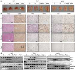
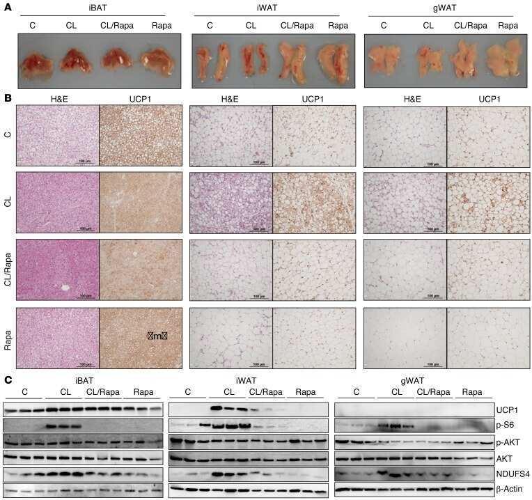
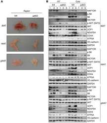
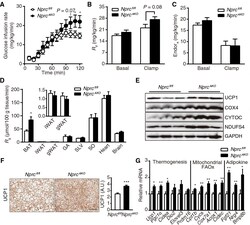
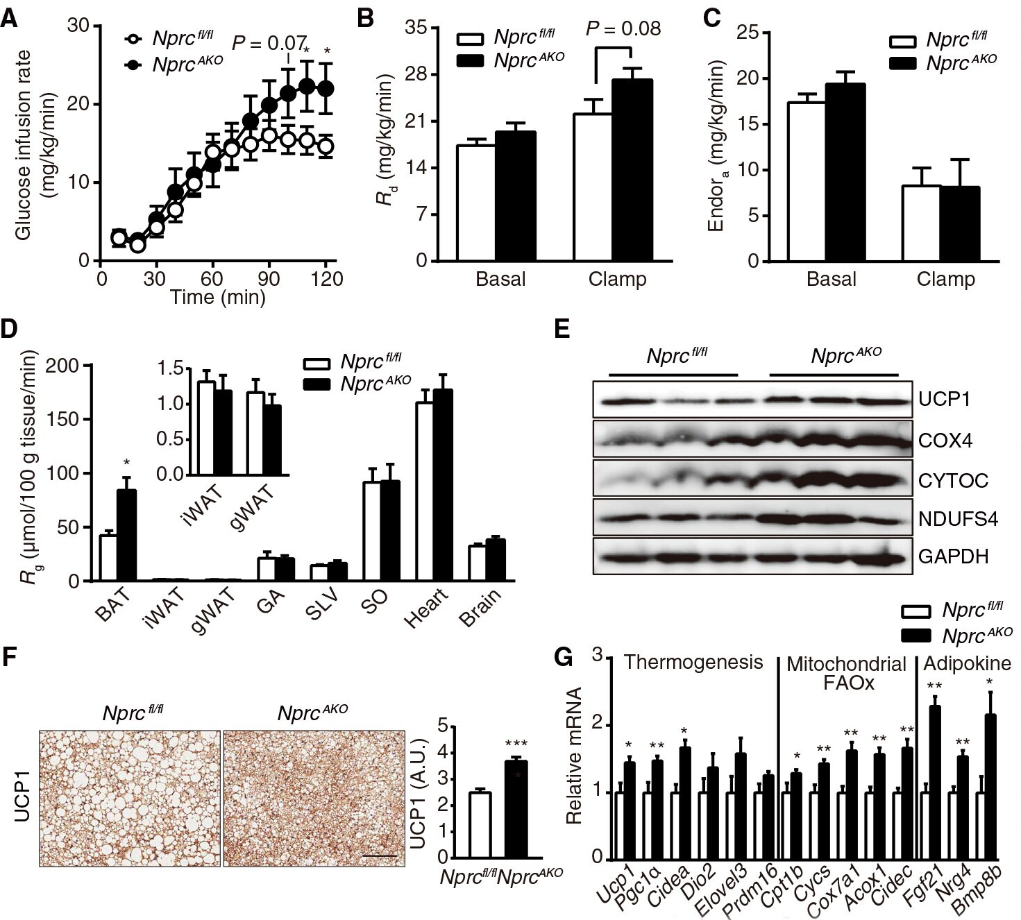
- Fig. 5. Glucose uptake and expression of thermogenesis markers are increased in the BAT of HFD-fed Nprc AKO mice. ( A ) Glucose infusion rate of Nprc AKO ( n = 10) and Nprc fl/fl ( n = 8) mice during a hyperinsulinemic-euglycemic clamp experiment performed at 12 weeks of HFD feeding. ( B and C ) Rates of glucose disposal ( R d ) (B) and endogenous glucose appearance (Endo R a ) (C) in Nprc AKO ( n = 10) and Nprc fl/fl ( n = 8) mice under basal and clamp states. ( D ) Rate of glucose uptake ( R g ) in the BAT, iWAT, gWAT, gastrocnemius (GA), superficial vastus lateralis (SVL), soleus (SO), heart, and brain of Nprc AKO ( n = 10) and Nprc fl/fl ( n = 8) mice at the end of the clamp experiment. ( E ) Western blot analysis of thermogenic and mitochondrial proteins in the BAT of Nprc AKO and Nprc fl/fl mice after 12 weeks on HFD. Blots are representative of three independent experiments. COX4, cytochrome c oxidase subunit 4; CYTOC, cytochrome c; NDUFS4, NADH (reduced form of nicotinamide adenine dinucleotide) dehydrogenase ubiquinone iron-sulfur protein 4; GAPDH, glyceraldehyde-3-phosphate dehydrogenase. ( F ) Immunostaining and quantification of UCP1 in the BAT of Nprc AKO ( n = 2) and Nprc fl/fl ( n = 2) mice after 12 weeks on HFD, as described in Materials and Methods. Scale bar, 200 mum. ( G ) qRT-PCR for the expression of genes coding thermogenic, mitochondrial, fatty acid oxidation (FAOx) markers, and BAT-derived adipokines in the BAT of Nprc AKO ( n = 14) and Nprc fl/fl ( n
- Submitted by
- Invitrogen Antibodies (provider)
- Main image

- Experimental details
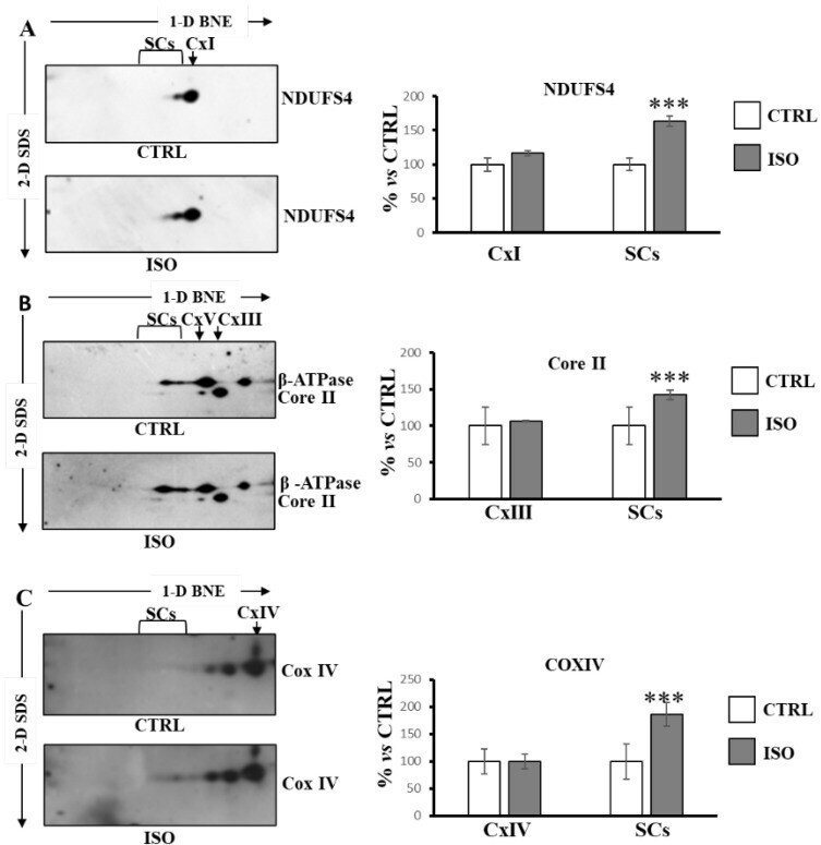
- Isoproterenol increased supercomplexes containing complex I, complex III and complex IV. The H9c2 cell cultures were incubated for 30 min in absence (CTRL) or in the presence of isoproterenol (ISO). Mitochondrial solubilized proteins were separated by 1D-BNE/2D-SDS-PAGE and transferred into nitrocellulose membrane, followed by immunoblotting analysis with specific antibodies. ( A ) Representative images of immunoblotting with antibody against NDUFS4. ( B ) Representative images of immunoblotting with antibodies against Core II and beta-ATP synthase. ( C ) Representative images of immunoblotting with antibody against Cox IV. ( A - C ) the histograms represent the percentage of ADU of isoproterenol-treated cells (ISO) with respect to untreated cells (CTRL) of immuno-revealed spots in free complexes (CxI, CxIII, CxIV) and in supercomplexes (SCs). The values are the means +- SD of three different experiments. (ISO vs. CTRL, *** p < 0.001, Student's t -test). For more details, see under Materials and Methods.