Explore
 Validate
 Learn
Learn Western blot
 ELISA
ELISAAntibody data
- Antibody Data
- Antigen structure
- References [0]
- Comments [0]
- Validations
- Western blot [1]
- Immunocytochemistry [1]
- Immunohistochemistry [1]
Submit
Validation data
Reference
Comment
Report error
- Product number
- GTX79211 - Provider product page

- Provider
- GeneTex
- Proper citation
- GeneTex Cat#GTX79211, RRID:AB_11179867
- Product name
- Recoverin antibody [6a55 cd6]
- Antibody type
- Monoclonal
- Reactivity
- Human, Rat, Bovine, Sheep
- Host
- Mouse
No comments: Submit comment
Supportive validation
- Submitted by
- GeneTex (provider)
- Main image
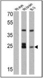
- Experimental details
- Western blot analysis of Recoverin in 25 ug of rat eye, rat brain and B-3 lysates. Proteins were transferred to a PVDF membrane and blocked at 4¢XC overnight. The membrane was probed with Recoverin antibody [6a55 cd6] at a dilution of 1:500 overnight at 4¢XC, washed in TBST, and probed with an HRP-conjugated secondary antibody. Chemiluminescent detection was performed.

Supportive validation
- Submitted by
- GeneTex (provider)
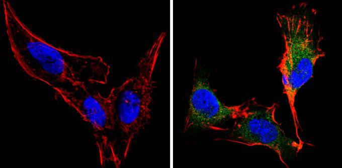
- Main image
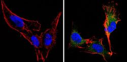
- Experimental details
- Immunofluorescent analysis of Recoverin (green) in B-3 cells (right) compared to a negative control without primary antibody (left). Formalin-fixed cells were permeabilized with 0.1% Triton X-100 in TBS for 5-10 minutes and blocked with 3% BSA-PBS for 30 minutes at room temperature. Cells were probed with Recoverin antibody [6a55 cd6] in 3% BSA-PBS at a dilution of 1:100 and incubated overnight at 4¢XC in a humidified chamber. Cells were washed with PBST and incubated with a proper secondary antibody. F-actin (red) was stained with a flourescent red phalloidin and nuclei (blue) were stained with Hoechst or DAPI. Images were taken at a magnification of 60x.
Supportive validation
- Submitted by
- GeneTex (provider)
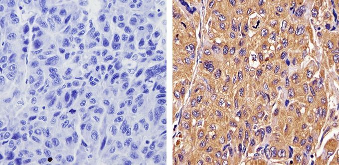
- Main image
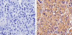
- Experimental details
- Immunohistochemistry analysis of Recoverin in paraffin-embedded human hepatocarcinoma (right) compared with a negative control in the absence of primary antibody (left). To expose target proteins, antigen retrieval was performed using 10mM sodium citrate (pH 6.0), microwaved for 8-15 min. Following antigen retrieval, tissues were blocked in 3% H2O2-methanol for 15 min at room temperature, washed with ddH2O and PBS, and then probed with Recoverin antibody [6a55 cd6] by 3% BSA-PBS at a dilution of 1:50 overnight at 4¢XC in a humidified chamber. Tissues were washed extensively in PBST and detection was performed using an HRP-conjugated secondary antibody followed by colorimetric detection using a DAB kit. Tissues were counterstained with hematoxylin and dehydrated with ethanol and xylene to prep for mounting.