Explore
 Validate
 Learn
Learn Western blot
Antibody data
- Antibody Data
- Antigen structure
- References [0]
- Comments [0]
- Validations
- Western blot [3]
- Immunocytochemistry [1]
- Immunohistochemistry [1]
Submit
Validation data
Reference
Comment
Report error
- Product number
- PA5-52011 - Provider product page

- Provider
- Invitrogen Antibodies
- Product name
- NDUFB8 Polyclonal Antibody
- Antibody type
- Polyclonal
- Antigen
- Recombinant full-length protein
- Description
- Immunogen sequence: GARTASHMTK DMFPGPYPRT PEERAAAAKK YNMRVEDYEP YPDDGMGYGD YPKLPDRSQH ERDPWYSWDQ PGLRLNWGEP MHWHLDM
- Concentration
- 0.3 mg/mL
No comments: Submit comment
Supportive validation
- Submitted by
- Invitrogen Antibodies (provider)
- Main image

- Experimental details
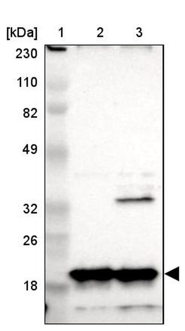
- Western blot analysis of NDUFB8 in Lane 1: Marker (kDa) 230, 110, 82, 49, 32, 26, 18; Lane 2: Human cell line RT-4; Lane 3: Human cell line U-251MG sp. Samples were probed using a NDUFB8 Polyclonal Antibody (Product # PA5-52011).
- Submitted by
- Invitrogen Antibodies (provider)
- Main image

- Experimental details
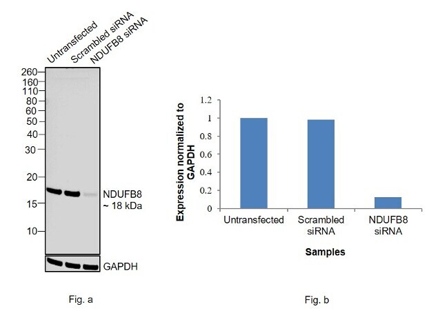
- Knockdown of NDUFB8 was achieved by transfecting A549 with NDUFB8 specific siRNAs (Silencer® select Product # s9368, s9369). Western blot analysis (Fig. a) was performed using membrane enriched extracts from the NDUFB8 knockdown cells (Lane 3), non-specific scrambled siRNA transfected cells (Lane 2) and untransfected cells (Lane 1). The blot was probed with NDUFB8 Polyclonal Antibody (Product # PA5-52011, 0.4 µg/mL) and Goat anti-Rabbit IgG (H+L) Superclonal™ Secondary Antibody, HRP conjugate (Product # A27036, 0.25 µg/mL, 1:4000 dilution). Densitometric analysis of this western blot is shown in histogram (Fig. b). Decrease in signal upon siRNA mediated knock down confirms that antibody is specific to NDUFB8.
- Submitted by
- Invitrogen Antibodies (provider)
- Main image

- Experimental details
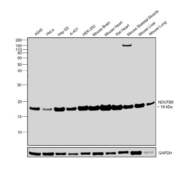
- Western blot was performed using Anti-NDUFB8 Polyclonal Antibody (Product # PA5-52011) and a 18 kDa band corresponding to NDUFB8 was observed across cell lines and tissue extracts tested. Membrane enriched extracts (30 µg lysate) of A549 (Lane 1), HeLa (Lane 2), Hep G2 (Lane 3), A-431 (Lane 4), HEK-293 (Lane 5) and tissue extracts (30ug lysate) of Mouse Brain (Lane 6), Mouse Heart (Lane 7), Rat Heart (Lane 8), Mouse Skeletal Muscle (Lane 9), Mouse Liver (Lane 10) and Mouse Lung (Lane 11) were electrophoresed using NuPAGE® 12 % Bis-Tris gel (Product # NP0342BOX). Resolved proteins were then transferred onto a nitrocellulose membrane (Product # IB23001) by dry transfer. The blot was probed with the primary antibody (0.4 ug/ml) and detected by chemiluminescence Goat Anti-Rabbit IgG Secondary Antibody, HRP conjugate (Product # A27036, 1:4000 dilution) using the iBright FL 1000 (Product # A32752). Chemiluminescent detection was performed using Novex® ECL Chemiluminescent Substrate Reagent Kit (Product # WP20005).
Supportive validation
- Submitted by
- Invitrogen Antibodies (provider)
- Main image
- Experimental details
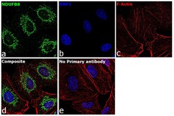
- Immunofluorescence analysis of NDUFB8 was performed using 70% confluent log phase A549 cells. The cells were fixed with 4% paraformaldehyde for 10 minutes, permeabilized with 0.1% Triton™ X-100 for 15 minutes, and blocked with 2% BSA for 1 hour at room temperature. The cells were labeled with NDUFB8 Rabbit Polyclonal Antibody (Product # PA5-52011) at 5 µg/mL in 0.1% BSA, incubated at 4 degree Celsius overnight and then labeled with Goat anti-Rabbit IgG (H+L) Superclonal™ Secondary Antibody, Alexa Fluor® 488 conjugate (Product # A27034) at a dilution of 1:2000 for 45 minutes at room temperature (Panel a: green). Nuclei (Panel b: blue) were stained with ProLong™ Diamond Antifade Mountant with DAPI (Product # P36962). F-actin (Panel c: red) was stained with Rhodamine Phalloidin (Product # R415). Panel d represents the merged image showing cytoplasmic localization. Panel e represents control cells with no primary antibody to assess background. The images were captured at 60X magnification.
Supportive validation
- Submitted by
- Invitrogen Antibodies (provider)
- Main image
- Experimental details
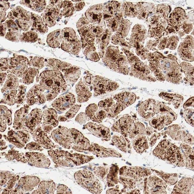
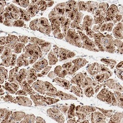
- Immunohistochemical staining of NDUFB8 in human heart muscle shows strong granular positivity in myocytes. Samples were probed using a NDUFB8 Polyclonal Antibody (Product # PA5-52011).