Explore
 Validate
 Learn
Learn Western blot
 Immunocytochemistry
Antibody data
- Antibody Data
- Antigen structure
- References [1]
- Comments [0]
- Validations
- Immunocytochemistry [1]
- Immunohistochemistry [1]
Submit
Validation data
Reference
Comment
Report error
- Product number
- HPA005640 - Provider product page

- Provider
- Atlas Antibodies
- Proper citation
- Atlas Antibodies Cat#HPA005640, RRID:AB_1079461
- Product name
- Anti-NDUFB5
- Antibody type
- Polyclonal
- Description
- Polyclonal Antibody against Human NDUFB5, Gene description: NADH dehydrogenase (ubiquinone) 1 beta subcomplex, 5, 16kDa, Alternative Gene Names: CI-SGDH, MGC12314, SGDH, Validated applications: ICC, IHC, WB, Uniprot ID: O43674, Storage: Store at +4°C for short term storage. Long time storage is recommended at -20°C.
- Reactivity
- Human
- Host
- Rabbit
- Conjugate
- Unconjugated
- Isotype
- IgG
- Vial size
- 100 µl
- Concentration
- 0.1 mg/ml
- Storage
- Store at +4°C for short term storage. Long time storage is recommended at -20°C.
- Handling
- The antibody solution should be gently mixed before use.
Submitted references p16Ink4a-induced senescence of pancreatic beta cells enhances insulin secretion
Helman A, Klochendler A, Azazmeh N, Gabai Y, Horwitz E, Anzi S, Swisa A, Condiotti R, Granit R, Nevo Y, Fixler Y, Shreibman D, Zamir A, Tornovsky-Babeay S, Dai C, Glaser B, Powers A, Shapiro A, Magnuson M, Dor Y, Ben-Porath I
Nature Medicine 2016;22(4):412-420
Nature Medicine 2016;22(4):412-420
No comments: Submit comment
Supportive validation
- Submitted by
- Atlas Antibodies (provider)
- Main image
- Experimental details
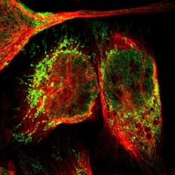
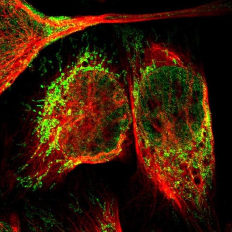
- Immunofluorescent staining of human cell line U-2 OS shows localization to nucleoplasm & mitochondria.
- Sample type
- Human
Supportive validation
- Submitted by
- Atlas Antibodies (provider)
- Enhanced method
- Orthogonal validation
- Main image

- Experimental details
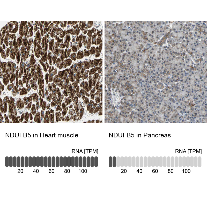
- Immunohistochemistry analysis in human heart muscle and pancreas tissues using HPA005640 antibody. Corresponding NDUFB5 RNA-seq data are presented for the same tissues.
- Sample type
- Human
- Protocol
- Protocol