Explore
 Validate
 Learn
LearnPA3-845
antibody from Invitrogen Antibodies
Targeting: IFIT2
cig42, G10P2, GARG-39, IFI-54, IFI54, ISG-54K
 Western blot
Antibody data
- Antibody Data
- Antigen structure
- References [7]
- Comments [0]
- Validations
- Western blot [1]
- Immunocytochemistry [4]
- Other assay [2]
Submit
Validation data
Reference
Comment
Report error
- Product number
- PA3-845 - Provider product page

- Provider
- Invitrogen Antibodies
- Product name
- IFIT2 Polyclonal Antibody
- Antibody type
- Polyclonal
- Antigen
- Other
- Description
- PA3-845 detects ISG54 (p54) from human and mouse samples. PA3-845 has been successfully used in ICC/IF and in Western blot applications to detect a 39-58 kDa band. This antibody may produce multiple background bands, and it is recommended to run samples slowly on an 8% gel to achieve proper separation.
- Reactivity
- Human, Mouse, Rat
- Host
- Rabbit
- Isotype
- IgG
- Vial size
- 100 µL
- Concentration
- Conc. Not Determined
- Storage
- -20° C, Avoid Freeze/Thaw Cycles
Submitted references MEK/ERK MAP kinase limits poly I:C-induced antiviral gene expression in RAW264.7 macrophages by reducing interferon-beta expression.
Butyrate Reprograms Expression of Specific Interferon-Stimulated Genes.
Interferon regulatory factor 3 plays a role in macrophage responses to interferon-γ.
Activation of IRF3 contributes to IFN-γ and ISG54 expression during the immune responses to B16F10 tumor growth.
Coronavirus nonstructural protein 15 mediates evasion of dsRNA sensors and limits apoptosis in macrophages.
Regional astrocyte IFN signaling restricts pathogenesis during neurotropic viral infection.
Type I IFNs Act upon Hematopoietic Progenitors To Protect and Maintain Hematopoiesis during Pneumocystis Lung Infection in Mice.
Freed SM, Baldi DS, Snow JA, Athen SR, Guinn ZP, Pinkerton TS, Petro TM, Moore TC
FEBS letters 2021 Nov;595(21):2665-2674
FEBS letters 2021 Nov;595(21):2665-2674
Butyrate Reprograms Expression of Specific Interferon-Stimulated Genes.
Chemudupati M, Kenney AD, Smith AC, Fillinger RJ, Zhang L, Zani A, Liu SL, Anderson MZ, Sharma A, Yount JS
Journal of virology 2020 Jul 30;94(16)
Journal of virology 2020 Jul 30;94(16)
Interferon regulatory factor 3 plays a role in macrophage responses to interferon-γ.
Guinn ZP, Petro TM
Immunobiology 2019 Jul;224(4):565-574
Immunobiology 2019 Jul;224(4):565-574
Activation of IRF3 contributes to IFN-γ and ISG54 expression during the immune responses to B16F10 tumor growth.
Guinn Z, Brown DM, Petro TM
International immunopharmacology 2017 Sep;50:121-129
International immunopharmacology 2017 Sep;50:121-129
Coronavirus nonstructural protein 15 mediates evasion of dsRNA sensors and limits apoptosis in macrophages.
Deng X, Hackbart M, Mettelman RC, O'Brien A, Mielech AM, Yi G, Kao CC, Baker SC
Proceedings of the National Academy of Sciences of the United States of America 2017 May 23;114(21):E4251-E4260
Proceedings of the National Academy of Sciences of the United States of America 2017 May 23;114(21):E4251-E4260
Regional astrocyte IFN signaling restricts pathogenesis during neurotropic viral infection.
Daniels BP, Jujjavarapu H, Durrant DM, Williams JL, Green RR, White JP, Lazear HM, Gale M Jr, Diamond MS, Klein RS
The Journal of clinical investigation 2017 Mar 1;127(3):843-856
The Journal of clinical investigation 2017 Mar 1;127(3):843-856
Type I IFNs Act upon Hematopoietic Progenitors To Protect and Maintain Hematopoiesis during Pneumocystis Lung Infection in Mice.
Prigge JR, Hoyt TR, Dobrinen E, Capecchi MR, Schmidt EE, Meissner N
Journal of immunology (Baltimore, Md. : 1950) 2015 Dec 1;195(11):5347-57
Journal of immunology (Baltimore, Md. : 1950) 2015 Dec 1;195(11):5347-57
No comments: Submit comment
Supportive validation
- Submitted by
- Invitrogen Antibodies (provider)
- Main image
- Experimental details
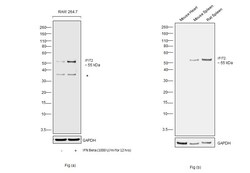
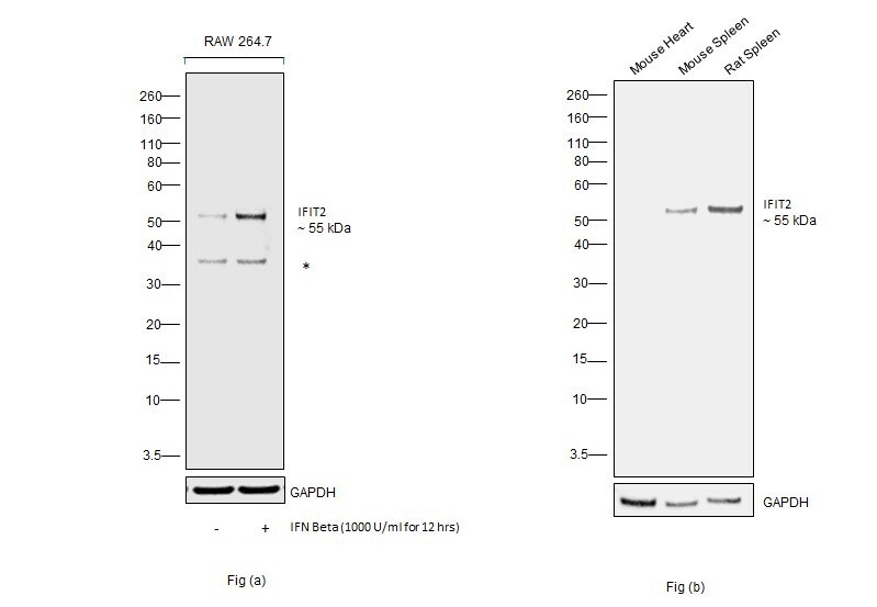
- Western blot was performed using Anti-IFIT2 Polyclonal Antibody (Product # PA3-845) and a 55 kDa band corresponding to Interferon-induced protein with tetratricopeptide repeats 2 was observed across cell lines and tissues tested. Whole cell extracts (30 µg lysate) of RAW 264.7 (Lane 1), RAW 264.7 treated with IFN Gamma (1000IU/mL for 12 hrs) (Lane 2) as seen in Fig (a) and Mouse Heart (Lane 1), Mouse Spleen (Lane 2) and Rat Spleen (Lane 3) as seen in Fig (b) were electrophoresed using NuPAGE™ 4-12% Bis-Tris Protein Gel (Product # NP0322BOX). Resolved proteins were then transferred onto a nitrocellulose membrane (Product # IB23002) by iBlot® 2 Dry Blotting System (Product # IB21001). The blot was probed with the primary antibody (1:1000 dilution) and detected by chemiluminescence with Goat anti-Rabbit IgG (H+L) Superclonal™ Recombinant Secondary Antibody, HRP (Product # A27036,1:4000 dilution) using the iBright FL 1000 (Product # A32752), an increased expression of IFIT2 was observed upon IFN Gamma treatment,. Relative expression of IFIT2 was observed to be high in Mouse Spleen in comparison to Mouse Heart. Chemiluminescent detection was performed using Novex® ECL Chemiluminescent Substrate Reagent Kit (Product # WP20005).
Supportive validation
- Submitted by
- Invitrogen Antibodies (provider)
- Main image
- Experimental details
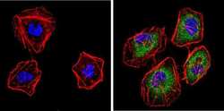
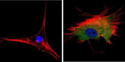
- Immunofluorescent analysis of p54 (green) showing staining in the cytoplasm of HeLa cells. Formalin-fixed cells were permeabilized with 0.1% Triton X-100 in TBS for 5-10 minutes and blocked with 3% BSA-PBS for 30 minutes at room temperature. Cells were probed with a p54 polyclonal antibody (Product # PA3-845) in 3% BSA-PBS at a dilution of 1:100 and incubated overnight at 4 ºC in a humidified chamber. Cells were washed with PBST and incubated with a DyLight-conjugated secondary antibody in PBS at room temperature in the dark. F-actin (red) was stained with a fluorescent red phalloidin and nuclei (blue) were stained with Hoechst or DAPI. Images were taken at a magnification of 60x.

- Submitted by
- Invitrogen Antibodies (provider)
- Main image
- Experimental details
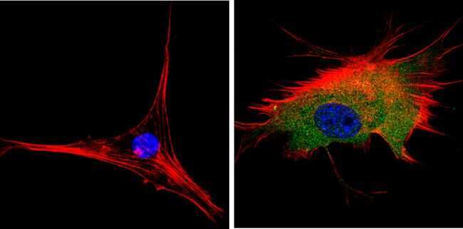
- Immunofluorescent analysis of p54 (green) showing staining in the cytoplasm of NIH-3T3 cells. Formalin-fixed cells were permeabilized with 0.1% Triton X-100 in TBS for 5-10 minutes and blocked with 3% BSA-PBS for 30 minutes at room temperature. Cells were probed with a p54 polyclonal antibody (Product # PA3-845) in 3% BSA-PBS at a dilution of 1:100 and incubated overnight at 4 ºC in a humidified chamber. Cells were washed with PBST and incubated with a DyLight-conjugated secondary antibody in PBS at room temperature in the dark. F-actin (red) was stained with a fluorescent red phalloidin and nuclei (blue) were stained with Hoechst or DAPI. Images were taken at a magnification of 60x.
- Submitted by
- Invitrogen Antibodies (provider)
- Main image
- Experimental details
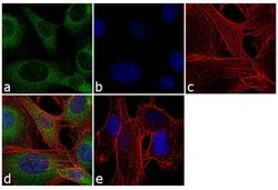
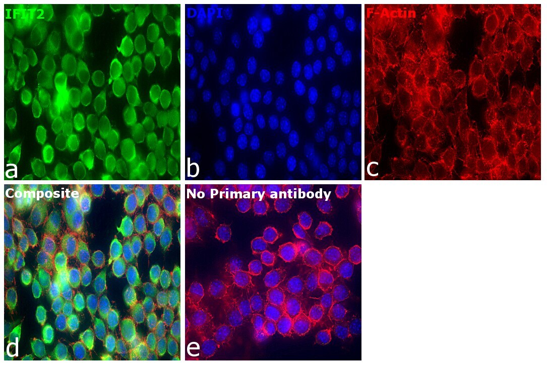
- Immunofluorescence analysis of IFIT2 was performed using 70% confluent log phase HT-1080 cells. The cells were fixed with 4% paraformaldehyde for 10 minutes, permeabilized with 0.1% Triton™ X-100 for 10 minutes, and blocked with 1% BSA for 1 hour at room temperature. The cells were labeled with IFIT2 Rabbit Polyclonal Antibody (Product # PA3-845) at 1:250 dilution in 0.1% BSA and incubated for 3 hours at room temperature and then labeled with Goat anti-Rabbit IgG (H+L) Superclonal™ Secondary Antibody, Alexa Fluor® 488 conjugate (Product # A27034) at a dilution of 1:2000 for 45 minutes at room temperature (Panel a: green). Nuclei (Panel b: blue) were stained with SlowFade® Gold Antifade Mountant with DAPI (Product # S36938). F-actin (Panel c: red) was stained with Rhodamine Phalloidin (Product # R415, 1:300). Panel d represents the merged image showing cytoplasmic localization. Panel e shows the no primary antibody control. The images were captured at 60X magnification.
- Submitted by
- Invitrogen Antibodies (provider)
- Main image

- Experimental details
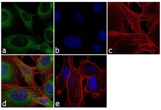
- Immunofluorescence analysis of Interferon-induced protein with tetratricopeptide repeats 2 was performed using 70% confluent log phase RAW 264.7 cells. The cells were fixed with 4% paraformaldehyde for 10 minutes, permeabilized with 0.1% Triton™ X-100 for 15 minutes, and blocked with 2% BSA for 45 minutes at room temperature. The cells were labeled with IFIT2 Polyclonal Antibody (Product # PA3-845) at 1:100 dilution n 0.1% BSA, incubated at 4 degree celsius overnight and then labeled with Donkey anti-Rabbit IgG (H+L) Highly Cross-Adsorbed Secondary Antibody, Alexa Fluor Plus 488 (Product # A32790), (1:2000 dilution), for 45 minutes at room temperature (Panel a: Green). Nuclei (Panel b:Blue) were stained with ProLong™ Diamond Antifade Mountant with DAPI (Product # P36962). F-actin (Panel c: Red) was stained with Rhodamine Phalloidin (Product # R415, 1:300 dilution). Panel d represents the merged image showing cytoplasmic localization. Panel e represents control cells with no primary antibody to assess background. The images were captured at 60X magnification.
Supportive validation
- Submitted by
- Invitrogen Antibodies (provider)
- Main image
- Experimental details
- NULL

- Submitted by
- Invitrogen Antibodies (provider)
- Main image

- Experimental details
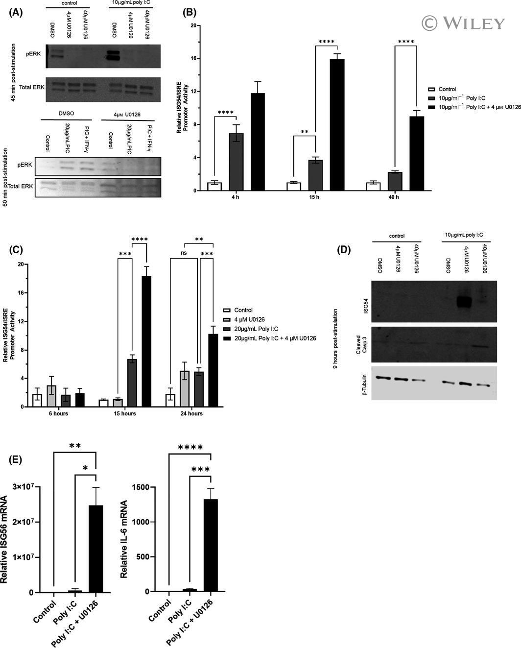
- 1 Fig. Blocking ERK with U0126 enhances poly I:C-induced activation of antiviral gene reporters. (A, D) RAW264.7 cells were seeded at 2 x 10 5 cells per well of a 24-well plate overnight before treating with or without 4 (A-E) or 40 u m (A, D) U0126 (or DMSO as a control). (A) After 45 min, cells were treated with 10 ug*mL -1 (top panel) or 20 ug*mL -1 (bottom panel) poly I:C or left untreated ('control'). Poly I:C + IFN-gamma was used as a positive control for ERK phosphorylation. 45 min (top panel) or 60 min (bottom panel) poststimulation with poly I:C, cells were lysed and analyzed by western blot (as described in the materials and methods) with anti-ERK p42/44 and anti-phosphorylated ERK. (B-C) RAW264.7-Lucia-ISG cells were seeded in 96-well plates for ISG54/ISRE luciferase reporter assay as described in the materials in methods. Briefly, cells were treated with 4 u m U0126 (or DMSO as a control) for 30 min prior to treatment with 10 ug*mL -1 (B) or 20 ug*mL -1 (C) poly I:C. At the time points indicated, supernatants were harvested and luciferase assays were performed as described in the materials and methods. (D) After 45 min, cells were treated with 10 ug*mL -1 poly I:C for 9 h. After incubation, cells were lysed and analyzed by western blot (as described in the materials and methods) with anti-ISG54, anticleaved caspase 3, and anti-beta tubulin. (E) 1x10 6 cells/well of a 6-well plate were seeded overnight in complete media. After overnight incubation, cells were pretr