Explore
 Validate
 Learn
Learn Western blot
 ELISA
ELISAAntibody data
- Antibody Data
- Antigen structure
- References [5]
- Comments [0]
- Validations
- Western blot [2]
Submit
Validation data
Reference
Comment
Report error
- Product number
- AF939 - Provider product page

- Provider
- R&D Systems
- Product name
- Human ADAM9 Ectodomain Antibody
- Antibody type
- Polyclonal
- Description
- Antigen Affinity-purified. Detects human ADAM9 Ectodomain in ELISAs and Western blots. In sandwich immunoassays, approximately 20% cross-reactivity with recombinant mouse (rm) rmADAM9 is observed and less than 0.05% cross-reactivity with recombinant human (rh) rhADAM8, rhADAM10, rhTACE, rhTIMP-1, rhTIMP-2, rhTIMP-3, rhTIMP-4, rhBACE-1 and rmADAM10 is observed.
- Reactivity
- Human
- Host
- Goat
- Conjugate
- Unconjugated
- Antigen sequence
Q13443- Isotype
- IgG
- Vial size
- 100 ug
- Concentration
- LYOPH
- Storage
- Use a manual defrost freezer and avoid repeated freeze-thaw cycles. 12 months from date of receipt, -20 to -70 °C as supplied. 1 month, 2 to 8 °C under sterile conditions after reconstitution. 6 months, -20 to -70 °C under sterile conditions after reconstitution.
Submitted references ADAM9 is over-expressed in human ovarian clear cell carcinomas and suppresses cisplatin-induced cell death.
Expression profiles and clinical correlations of degradome components in the tumor microenvironment of head and neck squamous cell carcinoma.
Role of ADAM-9 disintegrin-cysteine-rich domains in human keratinocyte migration.
Transmembrane collagen XVII, an epithelial adhesion protein, is shed from the cell surface by ADAMs.
Transmembrane collagen XVII, an epithelial adhesion protein, is shed from the cell surface by ADAMs.
Ueno M, Shiomi T, Mochizuki S, Chijiiwa M, Shimoda M, Kanai Y, Kataoka F, Hirasawa A, Susumu N, Aoki D, Okada Y
Cancer science 2018 Feb;109(2):471-482
Cancer science 2018 Feb;109(2):471-482
Expression profiles and clinical correlations of degradome components in the tumor microenvironment of head and neck squamous cell carcinoma.
Stokes A, Joutsa J, Ala-Aho R, Pitchers M, Pennington CJ, Martin C, Premachandra DJ, Okada Y, Peltonen J, Grénman R, James HA, Edwards DR, Kähäri VM
Clinical cancer research : an official journal of the American Association for Cancer Research 2010 Apr 1;16(7):2022-35
Clinical cancer research : an official journal of the American Association for Cancer Research 2010 Apr 1;16(7):2022-35
Role of ADAM-9 disintegrin-cysteine-rich domains in human keratinocyte migration.
Zigrino P, Steiger J, Fox JW, Löffek S, Schild A, Nischt R, Mauch C
The Journal of biological chemistry 2007 Oct 19;282(42):30785-93
The Journal of biological chemistry 2007 Oct 19;282(42):30785-93
Transmembrane collagen XVII, an epithelial adhesion protein, is shed from the cell surface by ADAMs.
Franzke CW, Tasanen K, Schäcke H, Zhou Z, Tryggvason K, Mauch C, Zigrino P, Sunnarborg S, Lee DC, Fahrenholz F, Bruckner-Tuderman L
The EMBO journal 2002 Oct 1;21(19):5026-35
The EMBO journal 2002 Oct 1;21(19):5026-35
No comments: Submit comment
Supportive validation
- Submitted by
- R&D Systems (provider)
- Main image
- Experimental details
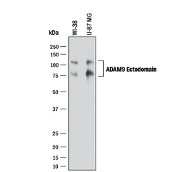
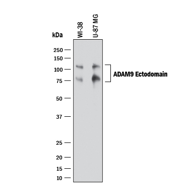
- Detection of Human ADAM9 by Western Blot. Western blot shows lysates of WI-38 human lung fibroblast cell line and U-87 MG human glioblastoma/astrocytoma cell line. PVDF membrane was probed with 1 µg/mL of Goat Anti-Human ADAM9 Ectodomain Antigen Affinity-purified Polyclonal Antibody (Catalog # AF939) followed by HRP-conjugated Anti-Goat IgG Secondary Antibody (Catalog # HAF017). Specific bands were detected for ADAM9 at approximately 110 and 80 kDa (as indicated). This experiment was conducted under reducing conditions and using Immunoblot Buffer Group 1.
- Submitted by
- R&D Systems (provider)
- Main image

- Experimental details
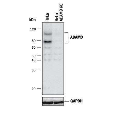
- Western Blot Shows Human ADAM9 Specificity by Using Knockout Cell Line. Western blot shows lysates of HeLa human cervical epithelial carcinoma parental cell line and ADAM9 knockout HeLa cell line (KO). PVDF membrane was probed with 1 µg/mL of Goat Anti-Human ADAM9 Ectodomain Antigen Affinity-purified Polyclonal Antibody (Catalog # AF939) followed by HRP-conjugated Anti-Sheep IgG Secondary Antibody (Catalog # HAF016). Specific bands were detected for ADAM9 at approximately 78 and 110 kDa (as indicated) in the parental HeLa cell line, but is not detectable in knockout HeLa cell line. GAPDH (Catalog # AF5718) is shown as a loading control. This experiment was conducted under reducing conditions and using Immunoblot Buffer Group 1.