Explore
 Validate
 Learn
Learn Western blot
 Immunocytochemistry
Antibody data
- Antibody Data
- Antigen structure
- References [2]
- Comments [0]
- Validations
- Western blot [3]
- Immunohistochemistry [7]
Submit
Validation data
Reference
Comment
Report error
- Product number
- NBP1-89345 - Provider product page

- Provider
- Novus Biologicals
- Proper citation
- Novus Cat#NBP1-89345, RRID:AB_11009652
- Product name
- Rabbit Polyclonal IFITM1 Antibody
- Antibody type
- Polyclonal
- Description
- Immunogen affinity purified. Specificity of human IFITM1 antibody verified on a Protein Array containing target protein plus 383 other non-specific proteins.
- Reactivity
- Human
- Host
- Rabbit
- Isotype
- IgG
- Vial size
- 0.1 ml
- Storage
- Store at 4C short term. Aliquot and store at -20C long term. Avoid freeze-thaw cycles.
Submitted references Targeted development of specific biomarkers of endometrial stromal cell differentiation using bioinformatics: the IFITM1 model.
In vivo functional requirement of the mouse Ifitm1 gene for germ cell development, interferon mediated immune response and somitogenesis.
Parra-Herran CE, Yuan L, Nucci MR, Quade BJ
Modern pathology : an official journal of the United States and Canadian Academy of Pathology, Inc 2014 Apr;27(4):569-79
Modern pathology : an official journal of the United States and Canadian Academy of Pathology, Inc 2014 Apr;27(4):569-79
In vivo functional requirement of the mouse Ifitm1 gene for germ cell development, interferon mediated immune response and somitogenesis.
Klymiuk I, Kenner L, Adler T, Busch DH, Boersma A, Irmler M, Fridrich B, Gailus-Durner V, Fuchs H, Leitner N, Müller M, Kühn R, Schlederer M, Treise I, de Angelis MH, Beckers J
PloS one 2012;7(10):e44609
PloS one 2012;7(10):e44609
No comments: Submit comment
Supportive validation
- Submitted by
- Novus Biologicals (provider)
- Main image

- Experimental details
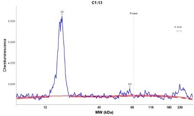
- Simple Western: IFITM1 Antibody [NBP1-89345] - Simple Western lane view shows a specific band for IFITM1 in 0.2 mg/ml of h. Tonsil lysate. This experiment was performed under reducing conditions using the 12-230 kDa separation system.

- Submitted by
- Novus Biologicals (provider)
- Main image
- Experimental details
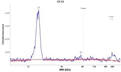
- Simple Western: IFITM1 Antibody [NBP1-89345] - Electropherogram image(s) of corresponding Simple Western lane view. IFITM1 antibody was used at 1:20 dilution on h. Tonsil lysate(s).
- Submitted by
- Novus Biologicals (provider)
- Main image
- Experimental details
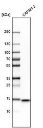
- Western Blot: IFITM1 Antibody [NBP1-89345] - Analysis in human cell line CAPAN-2.

Supportive validation
- Submitted by
- Novus Biologicals (provider)
- Main image
- Experimental details
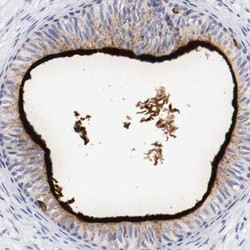
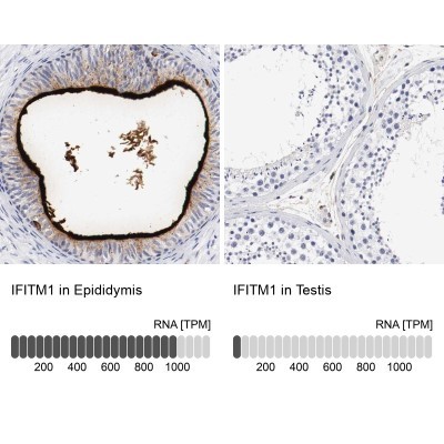
- Immunohistochemistry-Paraffin: IFITM1 Antibody [NBP1-89345] - Staining of human epididymis shows high expression.
- Submitted by
- Novus Biologicals (provider)
- Main image
- Experimental details
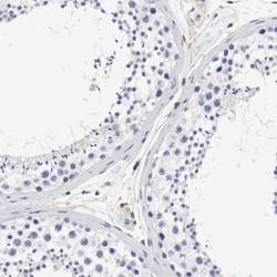
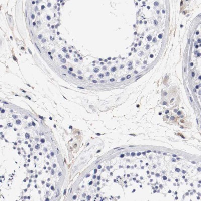
- Immunohistochemistry-Paraffin: IFITM1 Antibody [NBP1-89345] - Staining of human testis shows low expression as expected.
- Submitted by
- Novus Biologicals (provider)
- Main image
- Experimental details
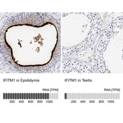
- Immunohistochemistry-Paraffin: IFITM1 Antibody [NBP1-89345] - Staining in human epididymis and testis tissues using anti-IFITM1 antibody. Corresponding IFITM1 RNA-seq data are presented for the same tissues.
- Submitted by
- Novus Biologicals (provider)
- Main image

- Experimental details
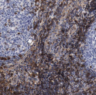
- Immunohistochemistry-Paraffin: IFITM1 Antibody [NBP1-89345] - Staining of human tonsil shows strong membranous positivity in non-germinal center cells.
- Submitted by
- Novus Biologicals (provider)
- Main image

- Experimental details
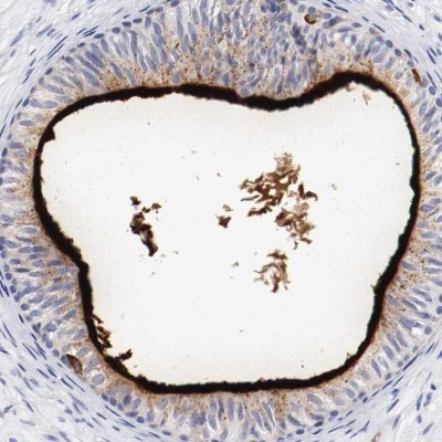
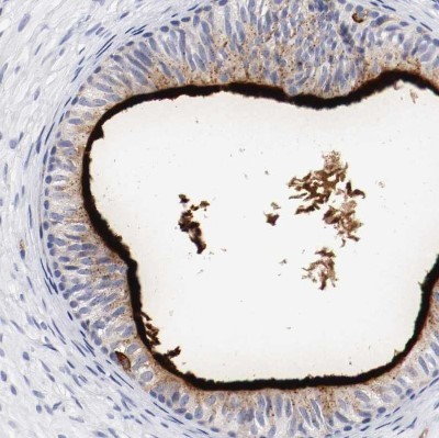
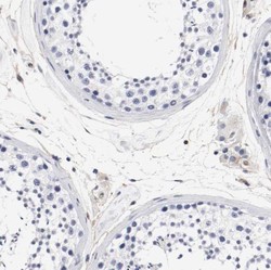
- Immunohistochemistry-Paraffin: IFITM1 Antibody [NBP1-89345] - Staining of human epididymis shows strong membranous positivity in ependymal cells.
- Submitted by
- Novus Biologicals (provider)
- Main image
- Experimental details
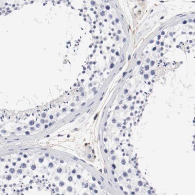
- Immunohistochemistry-Paraffin: IFITM1 Antibody [NBP1-89345] - Staining of human testis shows no membranous positivity in cells in seminiferous ducts as expected.
- Submitted by
- Novus Biologicals (provider)
- Main image
- Experimental details
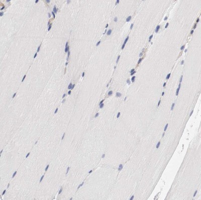
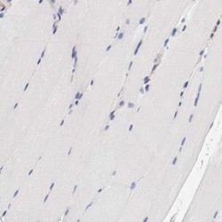
- Immunohistochemistry-Paraffin: IFITM1 Antibody [NBP1-89345] - Staining of human skeletal muscle shows no membranous positivity in myocytes as expected.