 Explore
 Validate
 Learn
Learn Western blot
 Immunocytochemistry
Antibody data
- Antibody Data
- Antigen structure
- References [1]
- Comments [0]
- Validations
- Western blot [5]
- Immunoprecipitation [1]
- Immunohistochemistry [3]
Submit
Validation data
Reference
Comment
Report error
- Product number
- NBP2-19711 - Provider product page

- Provider
- Novus Biologicals
- Product name
- Rabbit Polyclonal Pax6 Antibody
- Antibody type
- Polyclonal
- Description
- Immunogen affinity purified.
- Reactivity
- Human, Mouse, Rat
- Host
- Rabbit
- Isotype
- IgG
- Vial size
- 0.1 ml
- Storage
- Aliquot and store at -20C or -80C. Avoid freeze-thaw cycles.
Submitted references Production of neural stem cells from human pluripotent stem cells.
Wen Y, Jin S
Journal of biotechnology 2014 Oct 20;188:122-9
Journal of biotechnology 2014 Oct 20;188:122-9
No comments: Submit comment
Supportive validation
- Submitted by
- Novus Biologicals (provider)
- Main image

- Experimental details
- Western Blot: Pax6 Antibody [NBP2-19711] - Mouse tissue extract (50 ug) was separated by 10% SDS-PAGE, and the membrane was blotted with PAX6 antibody diluted at 1:1000. The HRP-conjugated anti-rabbit IgG antibody (NBP2-19301) was used to detect the primary antibody.

- Submitted by
- Novus Biologicals (provider)
- Main image

- Experimental details
- Western Blot: Pax6 Antibody [NBP2-19711] - Rat tissue extract (50 ug) was separated by 10% SDS-PAGE, and the membrane was blotted with PAX6 antibody diluted at 1:500. The HRP-conjugated anti-rabbit IgG antibody (NBP2-19301) was used to detect the primary antibody.

- Submitted by
- Novus Biologicals (provider)
- Main image

- Experimental details
- Western Blot: Pax6 Antibody [NBP2-19711] - Various whole cell extracts (30 ug) were separated by 10% SDS-PAGE, and the membrane was blotted with PAX6 antibody diluted by 1:1000.
- Submitted by
- Novus Biologicals (provider)
- Main image

- Experimental details
- Western Blot: Pax6 Antibody [NBP2-19711] - Various whole cell extracts (30 ug) were separated by 10% SDS-PAGE, and the membrane was blotted with PAX6 antibody diluted at 1:1000. The HRP-conjugated anti-rabbit IgG antibody (NBP2-19301) was used to detect the primary antibody.

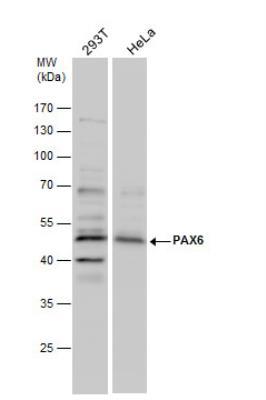
- Submitted by
- Novus Biologicals (provider)
- Main image

- Experimental details
- Western Blot: Pax6 Antibody [NBP2-19711] - Non-transfected (-) and transfected (+) 293T whole cell extracts (30 ug) were separated by 10% SDS-PAGE, and the membrane was blotted with PAX6 antibody diluted at 1:1000. The HRP-conjugated anti-rabbit IgG antibody (NBP2-19301) was used to detect the primary antibody.

Supportive validation
- Submitted by
- Novus Biologicals (provider)
- Main image
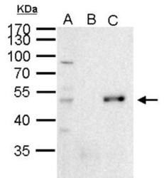
- Experimental details
- Immunoprecipitation: PAX6 Antibody [NBP2-19711] - Sample: 1000 ug 293T whole cell lysate/extract A. 50 ug 293T whole cell lysate/extract B. Control with 2 ug of preimmune rabbit IgG C. Immunoprecipitation of PAX6 protein by 2 ug of PAX6 antibody 10% SDS-PAGE The immunoprecipitated PAX6 protein was detected by PAX6 antibody diluted at 1:1000. anti-rabbit IgG was used as a secondary reagent.

Supportive validation
- Submitted by
- Novus Biologicals (provider)
- Main image
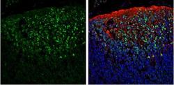
- Experimental details
- Immunohistochemistry-Frozen: Pax6 Antibody [NBP2-19711] - Frozen sectioned E13.5 Rat brain. Green: PAX6 protein stained by PAX6 antibody diluted at 1:250. Red: beta Tubulin 3/ TUJ1, a mature neuron marker, stained by beta Tubulin 3/ TUJ1 antibody [11710] (NBP2-43559) diluted at 1:500. Blue: Fluoroshield with DAPI.
- Submitted by
- Novus Biologicals (provider)
- Main image

- Experimental details
- Immunohistochemistry-Frozen: Pax6 Antibody [NBP2-19711] - Frozen section of embryonic mouse brain (mE12.5). PAX6 antibody diluted at 1:250.

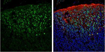
- Submitted by
- Novus Biologicals (provider)
- Main image

- Experimental details
- Immunohistochemistry-Paraffin: Pax6 Antibody [NBP2-19711] - Mouse retina. Green: PAX6 protein stained by PAX6 antibody diluted at 1:250. Red: beta Tubulin 3/ Tuj1, stained by beta Tubulin 3/ Tuj1 antibody [1338] diluted at 1:500. Blue: Fluoroshield with DAPI. Antigen Retrieval: Citrate buffer, pH 6.0, 15 min
