 Explore
 Validate
 Learn
Learn Western blot
Antibody data
- Antibody Data
- Antigen structure
- References [2]
- Comments [0]
- Validations
- Western blot [1]
- Immunocytochemistry [1]
- Flow cytometry [1]
Submit
Validation data
Reference
Comment
Report error
- Product number
- 720012 - Provider product page

- Provider
- Invitrogen Antibodies
- Product name
- Phospho-IRF3 (Ser396) Polyclonal Antibody
- Antibody type
- Polyclonal
- Antigen
- Synthetic peptide
- Description
- This antibody is predicted to react with Monkey.
- Reactivity
- Human
- Host
- Rabbit
- Isotype
- IgG
- Vial size
- 100 µg
- Concentration
- 0.5 mg/mL
- Storage
- Store at 4°C short term. For long term storage, store at -20°C, avoiding freeze/thaw cycles.
Submitted references The innate sensor ZBP1-IRF3 axis regulates cell proliferation in multiple myeloma.
Quercus acuta Thunb. (Fagaceae) and Its Component, Isoquercitrin, Inhibit HSV-1 Replication by Suppressing Virus-Induced ROS Production and NF-κB Activation.
Ponnusamy K, Tzioni MM, Begum M, Robinson ME, Caputo VS, Katsarou A, Trasanidis N, Xiao X, Kostopoulos IV, Iskander D, Roberts I, Trivedi P, Auner HW, Naresh K, Chaidos A, Karadimitris A
Haematologica 2022 Mar 1;107(3):721-732
Haematologica 2022 Mar 1;107(3):721-732
Quercus acuta Thunb. (Fagaceae) and Its Component, Isoquercitrin, Inhibit HSV-1 Replication by Suppressing Virus-Induced ROS Production and NF-κB Activation.
Kim B, Kim YS, Hwang YH, Yang HJ, Li W, Kwon EB, Kim TI, Go Y, Choi JG
Antioxidants (Basel, Switzerland) 2021 Oct 18;10(10)
Antioxidants (Basel, Switzerland) 2021 Oct 18;10(10)
No comments: Submit comment
Supportive validation
- Submitted by
- Invitrogen Antibodies (provider)
- Main image
- Experimental details
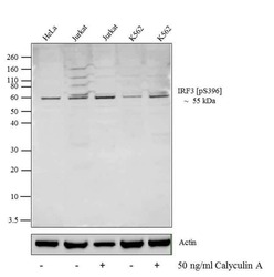
- Western blot analysis was performed on whole cell extracts (20 µg lysate) of HeLa (lane1), Jurkat (lane2), Jurkat treated Calyculin A (50 ng/mL/20 min) (lane3), K562 (lane4) and K562 treated with Calyculin A (50 ng/mL/20 min) (lane5). The blots were probed with Anti-IRF3 (pS396)Rabbit Polyclonal Antibody (Product # 720012, 0.5-2 µg/mL) and detected by chemiluminescence using Goat anti-Rabbit IgG (H+L) Superclonal Secondary Antibody, HRP conjugate (Product # A27036, 0.4 µg/mL, 1:2500 dilution). A 55 kDa band corresponding to IRF3 (pS396) was observed across cell lines tested according to the treatment. Known quantity of protein samples were electrophoresed using Novex®NuPAGE®4-12% Bis-Tris gel (Product # NP0321BOX), XCell SureLock Electrophoresis System (Product # EI0002), and Novex® Sharp Pre-Stained Protein Standard (Product # LC5800). Resolved proteins were then transferred onto a nitrocellulose membrane with iBlot® Dry Blotting System (Product # IB21001). The membrane was probed with the relevant primary and secondary antibody following blocking with 5% skimmed milk. Chemiluminescent detection was performed using Pierce™ ECL Western Blotting Substrate (Product # 32106).

Supportive validation
- Submitted by
- Invitrogen Antibodies (provider)
- Main image

- Experimental details
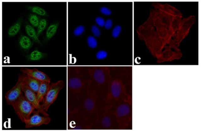
- Immunofluorescence was performed on fixed and permeabilized HeLa cells treated with Calyculin A (50 ng/mL/20 min) for detection of IRF-3 (pS396) using Anti-IRF-3 (pS396)Rabbit Polyclonal Antibody (Product # 720012, 2 µg/mL) and labeled with Goat anti-Rabbit IgG (H+L) Superclonal Secondary Antibody, Alexa Fluor® 488 conjugate (Product # A27034, 0.4 µg/mL, 1:2500). Panel a) shows representative cells that were stained for detection and localization of IRF-3 (pS396) protein (green), Panel b) is stained for nuclei (blue) using SlowFade® Gold Antifade Mountant with DAPI (Product # S36938, 1:50). Panel c) represents cytoskeletal F-actin staining using Alexa Fluor® 594 Phalloidin (Product # A12381, 1:200). Panel d) is a composite image of Panels a, b and c clearly demonstrating nuclear localization of IRF-3 (pS396). Panel e) represents control cells with no primary antibody to assess background.
Supportive validation
- Submitted by
- Invitrogen Antibodies (provider)
- Main image

- Experimental details
- Flow Cytometry analysis of IRF-3 [pS396] was performed on Jurkat cells treated with Calyculin A (50ng/ml/ 20 min) cells labeled with IRF-3 [pS396] Rabbit Polyclonal Antibody (Product# 720012, 2-4 ug/ 1M cells) or with rabbit isotype control and detected with Goat anti-Rabbit IgG (H+L) Superclonalª Secondary Antibody, Alexa Fluor¨ 488 conjugate (Product # A27034, 0.4 ug/ml, 1:2500) as represented by the red and pink histograms respectively. The purple histogram represents unstained control cells and the green histogram represents no-primary-antibody control. A representative 10,000 cells were acquired and analyzed for each sample using an Attune¨ Acoustic Focusing Cytometer (4468770).
