 Explore
 Validate
 Learn
Learn Western blot
Antibody data
- Antibody Data
- Antigen structure
- References [4]
- Comments [0]
- Validations
- Western blot [3]
- Other assay [2]
Submit
Validation data
Reference
Comment
Report error
- Product number
- 14-9947-82 - Provider product page

- Provider
- Invitrogen Antibodies
- Product name
- IRF3 Monoclonal Antibody (SL-12 (SL12)), eBioscience™
- Antibody type
- Monoclonal
- Antigen
- Other
- Description
- Description: This SL-12 monoclonal antibody reacts with human Interferon Regulatory Factor (IRF)-3. IRF-3 is a 427 amino acid constitutively expressed member of the IRF transcription factor family that plays a critical role in the induction of type I interferons. Inactive IRF-3 continuously transits between the cytoplasm and nucleus. Virus or dsRNA induces the C-terminal phosphorylation of IRF-3 by IKKε and TBK-1. This phosphorylation results in retention of IRF-3 in the nucleus and subsequent transcription of IFN-β, several interferon stimulated genes (ISGs), and other antiviral response genes.
- Antibody clone number
- SL-12 (SL12)
- Concentration
- 0.5 mg/mL
Submitted references Triggering the innate antiviral response through IRF-3 activation.
Recruitment of activated IRF-3 and CBP/p300 to herpes simplex virus ICP0 nuclear foci: Potential role in blocking IFN-beta induction.
Essential role of interferon regulatory factor 3 in direct activation of RANTES chemokine transcription.
Identification of a member of the interferon regulatory factor family that binds to the interferon-stimulated response element and activates expression of interferon-induced genes.
Hiscott J
The Journal of biological chemistry 2007 May 25;282(21):15325-9
The Journal of biological chemistry 2007 May 25;282(21):15325-9
Recruitment of activated IRF-3 and CBP/p300 to herpes simplex virus ICP0 nuclear foci: Potential role in blocking IFN-beta induction.
Melroe GT, Silva L, Schaffer PA, Knipe DM
Virology 2007 Apr 10;360(2):305-21
Virology 2007 Apr 10;360(2):305-21
Essential role of interferon regulatory factor 3 in direct activation of RANTES chemokine transcription.
Lin R, Heylbroeck C, Genin P, Pitha PM, Hiscott J
Molecular and cellular biology 1999 Feb;19(2):959-66
Molecular and cellular biology 1999 Feb;19(2):959-66
Identification of a member of the interferon regulatory factor family that binds to the interferon-stimulated response element and activates expression of interferon-induced genes.
Au WC, Moore PA, Lowther W, Juang YT, Pitha PM
Proceedings of the National Academy of Sciences of the United States of America 1995 Dec 5;92(25):11657-61
Proceedings of the National Academy of Sciences of the United States of America 1995 Dec 5;92(25):11657-61
No comments: Submit comment
Supportive validation
- Submitted by
- Invitrogen Antibodies (provider)
- Main image

- Experimental details
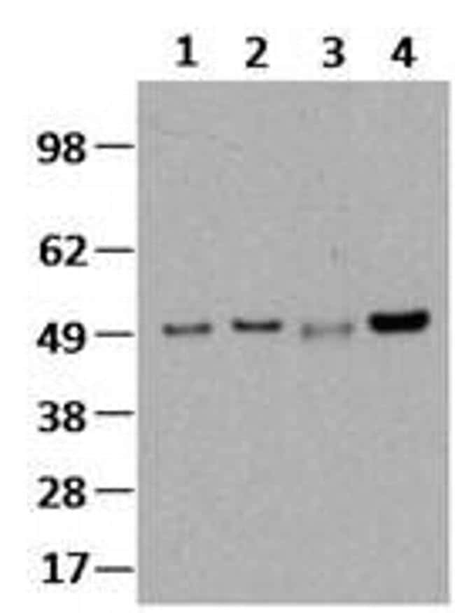
- Lysates prepared from Jurkat cells (lanes 1 and 2) and PBMCs (lanes 3 and 4) under nonreducing conditions (lanes 1 and 3) or reduced with DTT (lanes 2 and 4) were resolved by SDS-PAGE then immunoblotted with 2 µg/mL of Anti-Human IRF3 Purified. Bands were visualized using Anti-Mouse IgG HRP.
- Submitted by
- Invitrogen Antibodies (provider)
- Main image

- Experimental details
- Knockdown of IRF3 was achieved by transfecting A549 with IRF3 specific siRNAs (Silencer® select Product # s7509, s7507). Western blot analysis (Fig. a) was performed using whole cell extracts from the IRF3 knockdown cells (lane 3), non-specific scrambled siRNA transfected cells (lane 2) and untransfected cells (lane 1). The blot was probed with IRF3 Monoclonal Antibody (SL-12 (SL12)), eBioscience™ (Product # 14-9947-82, 2ug/ml) and Goat anti-Mouse IgG (H+L), Superclonal™ Recombinant Secondary Antibody, HRP (Product # A28177, 0.25µg/ml, 1:4000 dilution). Densitometric analysis of this western blot is shown in histogram (Fig. b). Decrease in signal upon siRNA mediated knock down confirms that antibody is specific to IRF3.

- Submitted by
- Invitrogen Antibodies (provider)
- Main image

- Experimental details
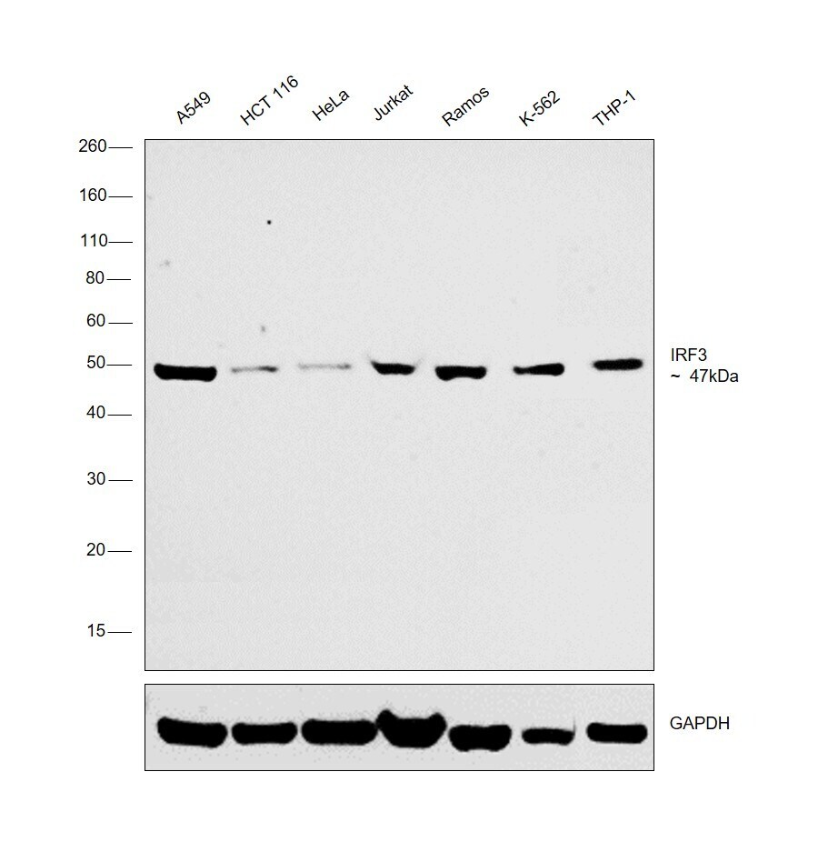
- Western blot was performed using Anti-IRF3 Monoclonal Antibody (SL-12 (SL12)), eBioscience™ (Product # 14-9947-82) and a 47 kDa band corresponding to IRF3 was observed across cell lines tested. Modified whole cell extracts (1% SDS) (30 µg lysate) of A549 (Lane 1), HCT 116 (Lane 2), HeLa (Lane 3), Jurkat (Lane 4), Ramos (Lane 5), K-562 (Lane 6) and THP-1 (Lane 7) were electrophoresed using NuPAGE™ 4-12% Bis-Tris Protein Gel (Product # NP0322BOX). Resolved proteins were then transferred onto a nitrocellulose membrane (Product # IB23001) by iBlot® 2 Dry Blotting System (Product # IB21001). The blot was probed with the primary antibody (2ug/ml) and detected by chemiluminescence with Goat anti-Mouse IgG (H+L) Superclonal™ Recombinant Secondary Antibody, HRP (Product # A28177, 1:4000 dilution) using the iBright FL 1000 (Product # A32752). Chemiluminescent detection was performed using Novex® ECL Chemiluminescent Substrate Reagent Kit (Product # WP20005).
Supportive validation
- Submitted by
- Invitrogen Antibodies (provider)
- Main image

- Experimental details
- NULL
- Submitted by
- Invitrogen Antibodies (provider)
- Main image
- Experimental details
- NULL
