Explore
 Validate
 Learn
Learn Western blot
Antibody data
- Antibody Data
- Antigen structure
- References [0]
- Comments [0]
- Validations
- Western blot [2]
- Immunocytochemistry [2]
- Flow cytometry [5]
- Protein array [1]
- Other assay [1]
Submit
Validation data
Reference
Comment
Report error
- Product number
- V9201 - Provider product page

- Provider
- NSJ Bioreagents
- Product name
- IRF3 Antibody / Interferon regulatory factor 3
- Antibody type
- Monoclonal
- Description
- This highly specific IRF3 antibody is suitable for use in Flow cytometry/Immunofluorescence/Western blot applications with human samples.
- Reactivity
- Human
- Host
- Mouse
- Conjugate
- Unconjugated
- Antibody clone number
- PCRP-IRF3-6C8
- Vial size
- 20 ug (with BSA and sodium azide), 100 ug (with BSA and sodium azide), 100 ug (without BSA or sodium azide), 7 ml IHC only format (if applicable)
- Concentration
- 0.2 mg/ml, 1 mg/ml
- Storage
- Aliquot the IRF3 antibody and store frozen at -20oC or colder. Avoid repeated freeze-thaw cycles.
No comments: Submit comment
Supportive validation
- Submitted by
- NSJ Bioreagents (provider)
- Main image
- Experimental details
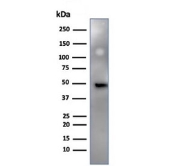
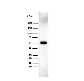
- Western blot testing of human HeLa cell lysate using IRF3 antibody (clone PCRP-IRF3-6C8). Predicted molecular weight ~47 kDa.
- Submitted by
- NSJ Bioreagents (provider)
- Main image
- Experimental details
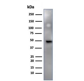
- Western blot testing of human MCF7 cell lysate using IRF3 antibody (clone PCRP-IRF3-6C8). Predicted molecular weight ~47 kDa.
Supportive validation
- Submitted by
- NSJ Bioreagents (provider)
- Main image
- Experimental details
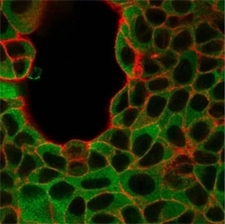
- Immunofluorescent staining of human MCF-7 cells using IRF3 antibody (green, clone PCRP-IRF3-6C8) and phalloidin (red).

- Submitted by
- NSJ Bioreagents (provider)
- Main image

- Experimental details
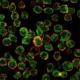
- Immunofluorescent staining of human K562 cells using IRF3 antibody (green, clone PCRP-IRF3-6C8) and phalloidin (red).
Supportive validation
- Submitted by
- NSJ Bioreagents (provider)
- Main image
- Experimental details
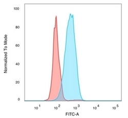
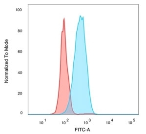
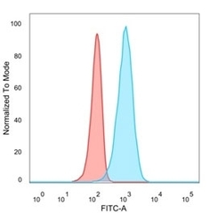
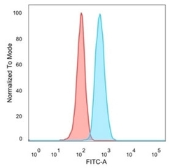
- FACS staining of PFA-fixed human MCF-7 cells with IRF3 antibody (blue, clone PCRP-IRF3-6C8), and unstained cells (red).
- Submitted by
- NSJ Bioreagents (provider)
- Main image
- Experimental details
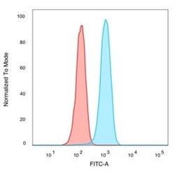
- FACS staining of PFA-fixed human K562 cells with IRF3 antibody (blue, clone PCRP-IRF3-6C8), and unstained cells (red).

- Submitted by
- NSJ Bioreagents (provider)
- Main image
- Experimental details
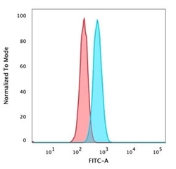
- FACS staining of PFA-fixed human HeLa cells with IRF3 antibody (blue, clone PCRP-IRF3-6C8), and unstained cells (red).
- Submitted by
- NSJ Bioreagents (provider)
- Main image
- Experimental details
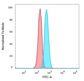
- FACS staining of PFA-fixed human U87 cells with IRF3 antibody (blue, clone PCRP-IRF3-6C8), and unstained cells (red).

- Submitted by
- NSJ Bioreagents (provider)
- Main image
- Experimental details
- FACS staining of PFA-fixed human Raji cells with IRF3 antibody (blue, clone PCRP-IRF3-6C8), and unstained cells (red).

Supportive validation
- Submitted by
- NSJ Bioreagents (provider)
- Main image

- Experimental details
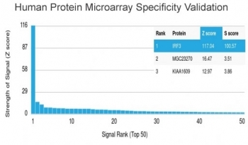
- Analysis of HuProt(TM) microarray containing more than 19,000 full-length human proteins using IRF3 antibody (clone PCRP-IRF3-6C8). These results demonstrate the foremost specificity of the PCRP-IRF3-6C8 mAb. Z- and S- score: The Z-score represents the strength of a signal that an antibody (in combination with a fluorescently-tagged anti-IgG secondary Ab) produces when binding to a particular protein on the HuProt(TM) array. Z-scores are described in units of standard deviations (SD's) above the mean value of all signals generated on that array. If the targets on the HuProt(TM) are arranged in descending order of the Z-score, the S-score is the difference (also in units of SD's) between the Z-scores. The S-score therefore represents the relative target specificity of an Ab to its intended target.
Supportive validation
- Submitted by
- NSJ Bioreagents (provider)
- Main image
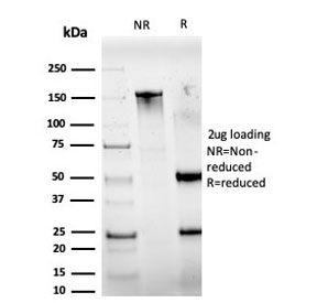
- Experimental details
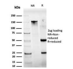
- SDS-PAGE analysis of purified, BSA-free IRF3 antibody (PCRP-IRF3-6C8) as confirmation of integrity and purity.