Explore
 Validate
 Learn
Learn Western blot
 ELISA
ELISAAntibody data
- Antibody Data
- Antigen structure
- References [0]
- Comments [0]
- Validations
- Western blot [1]
- Immunocytochemistry [1]
- Blocking/Neutralizing [1]
Submit
Validation data
Reference
Comment
Report error
- Product number
- AF2548 - Provider product page

- Provider
- R&D Systems
- Product name
- Human/Rhesus Macaque IL-18/IL-1F4 Antibody
- Antibody type
- Polyclonal
- Description
- Antigen Affinity-purified. Detects rhesus macaque IL-18/IL-1F4 in direct ELISAs and Western blots. In Western blots, approximately 10% cross-reactivity with recombinant mouse IL-18, recombinant rat IL-18, and recombinant porcine IL-18 is observed.
- Reactivity
- Human
- Host
- Goat
- Conjugate
- Unconjugated
- Antigen sequence
AAK13416- Isotype
- IgG
- Vial size
- 100 ug
- Concentration
- LYOPH
- Storage
- Use a manual defrost freezer and avoid repeated freeze-thaw cycles. 12 months from date of receipt, -20 to -70 °C as supplied. 1 month, 2 to 8 °C under sterile conditions after reconstitution. 6 months, -20 to -70 °C under sterile conditions after reconstitution.
No comments: Submit comment
Supportive validation
- Submitted by
- R&D Systems (provider)
- Main image
- Experimental details
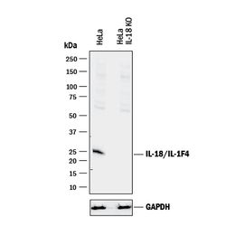
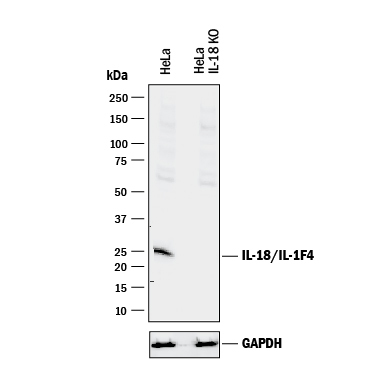
- Western Blot Shows Human IL-18/IL-1F4 Specificity by Using Knockout Cell Line. Western blot shows lysates of HeLa human cervical epithelial carcinoma parental cell line and IL-18/IL-1F4 knockout HeLa cell line (KO). PVDF membrane was probed with 1 µg/mL of Goat Anti-Human/Rhesus Macaque IL-18/IL-1F4 Antigen Affinity-purified Polyclonal Antibody (Catalog # AF2548) followed by HRP-conjugated Anti-Goat IgG Secondary Antibody (Catalog # HAF017). A specific band was detected for IL-18/IL-1F4 at approximately 25 kDa (as indicated) in the parental HeLa cell line, but is not detectable in knockout HeLa cell line. GAPDH (Catalog # AF5718) is shown as a loading control. This experiment was conducted under reducing conditions and using Immunoblot Buffer Group 1.
Supportive validation
- Submitted by
- R&D Systems (provider)
- Main image
- Experimental details
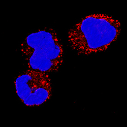
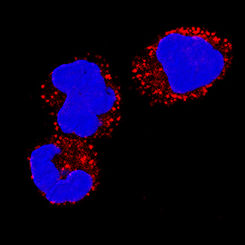
- IL-18/IL-1F4 in Human PBMCs. IL-18/IL-1F4 was detected in immersion fixed human peripheral blood mononuclear cells (PBMCs) using Goat Anti-Human/Rhesus Macaque IL-18/IL-1F4 Antigen Affinity-purified Polyclonal Antibody (Catalog # AF2548) at 5 µg/mL for 3 hours at room temperature. Cells were stained using the NorthernLights™ 557-conjugated Anti-Goat IgG Secondary Antibody (red; Catalog # NL001) and counterstained with DAPI (green). Specific staining was localized to cytoplasm. View our protocol for Fluorescent ICC Staining of Non-adherent Cells.
Supportive validation
- Submitted by
- R&D Systems (provider)
- Main image
- Experimental details
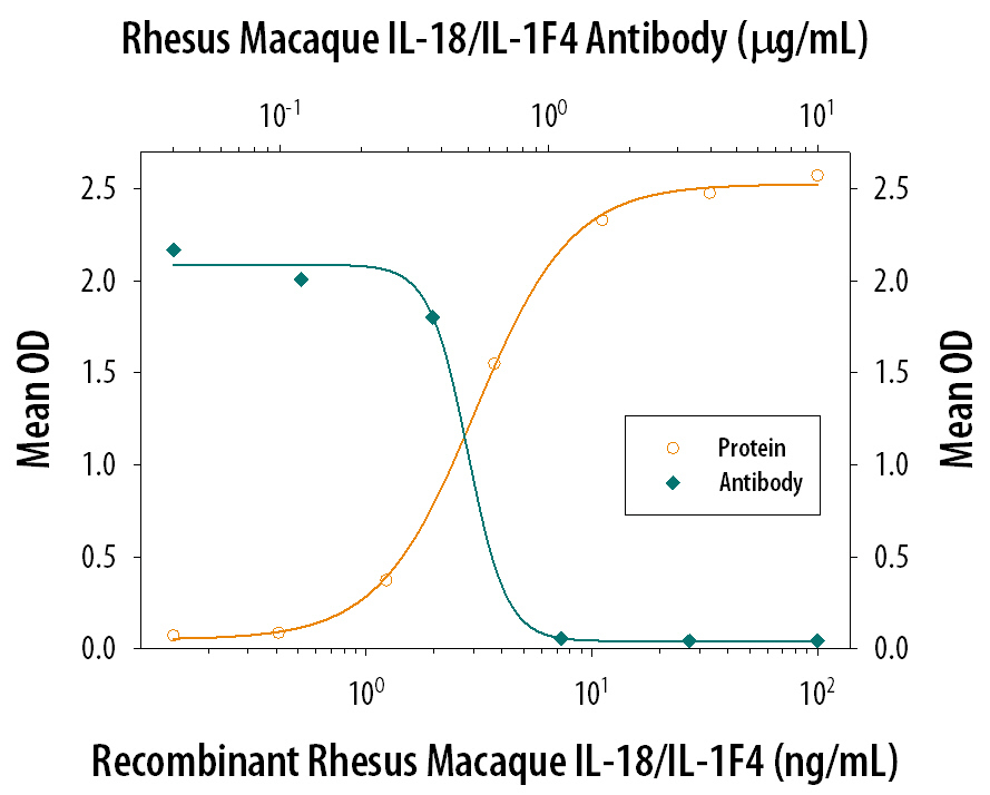
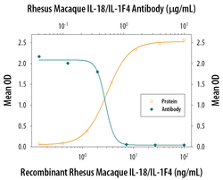
- IFN-gamma Secretion Induced by IL-18/IL-1F4 and Neutralization by Primate IL-18/IL-1F4 Antibody. In the presence of Recombinant Human TNF-alpha (20 ng/mL, Catalog # 210-TA), Recombinant Rhesus Macaque IL-18/IL-1F4 (Catalog # 2548-RM) stimulates IFN-gamma secretion in the KG-1 human acute myelogenous leukemia cell line in a dose-dependent manner (orange line), as measured by the Human IFN-gamma Quantikine ELISA Kit (Catalog # DIF50). Under these conditions, IFN-gamma secretion elicited by Recombinant Rhesus Macaque IL-18/IL-1F4 (10 ng/mL) is neutralized (green line) by increasing concentrations of Goat Anti-Human/Rhesus Macaque IL-18/IL-1F4 Antigen Affinity-purified Polyclonal Antibody (Catalog # AF2548). The ND50 is typically 0.4-1.2 µg/mL.