 Explore
 Validate
 Learn
Learn ELISA
ELISAAntibody data
- Antibody Data
- Antigen structure
- References [0]
- Comments [0]
- Validations
- ELISA [1]
- Immunocytochemistry [1]
- Flow cytometry [1]
Submit
Validation data
Reference
Comment
Report error
- Product number
- MAB2548-100 - Provider product page

- Provider
- R&D Systems
- Product name
- Human IL-18/IL-1F4 Antibody
- Antibody type
- Monoclonal
- Description
- Protein A or G purified from hybridoma culture supernatant. Detects human IL-18/IL-1F4 in direct ELISAs.
- Reactivity
- Human
- Host
- Mouse
- Conjugate
- Unconjugated
- Antigen sequence
Q14116- Isotype
- IgG
- Antibody clone number
- 925008
- Vial size
- 100 ug
- Storage
- Use a manual defrost freezer and avoid repeated freeze-thaw cycles. 12 months from date of receipt, -20 to -70 °C as supplied. 1 month, 2 to 8 °C under sterile conditions after reconstitution. 6 months, -20 to -70 °C under sterile conditions after reconstitution.
No comments: Submit comment
Supportive validation
- Submitted by
- R&D Systems (provider)
- Main image

- Experimental details
- Human IL-18/IL-1F4 ELISA Standard Curve. Recombinant Human IL-18/IL-1F4 protein was serially diluted 2-fold and captured by Rabbit Anti-Human IL-18/IL-1F4 Monoclonal Antibody (Catalog # MAB91243) coated on a Clear Polystyrene Microplate (Catalog # DY990). Mouse Anti-Human IL-18/IL-1F4 Monoclonal Antibody (Catalog # MAB2548) was biotinylated and incubated with the protein captured on the plate. Detection of the standard curve was achieved by incubating Streptavidin-HRP (Catalog # DY998) followed by Substrate Solution (Catalog # DY999) and stopping the enzymatic reaction with Stop Solution (Catalog # DY994).

Supportive validation
- Submitted by
- R&D Systems (provider)
- Main image
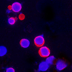
- Experimental details
- IL-18/IL-1F4 in Mouse Splenocytes. IL-18/IL-1F4 was detected in immersion fixed mouse splenocytes stimulated with Cal and PMA using Mouse Anti-Human IL-18/IL-1F4 Monoclonal Antibody (Catalog # MAB2548) at 25 µg/mL for 3 hours at room temperature. Cells were stained using the NorthernLights™ 557-conjugated Anti-Mouse IgG Secondary Antibody (red; Catalog # NL007) and counterstained with DAPI (blue). Specific staining was localized to cytoplasm. View our protocol for Fluorescent ICC Staining of Non-adherent Cells.

Supportive validation
- Submitted by
- R&D Systems (provider)
- Main image
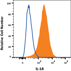
- Experimental details
- Detection of IL-18/IL-1F4 in Human THP-1 cell line by Flow Cytometry. THP-1 Human acute monocytic leukemia cell line was stained with Mouse Anti-Human IL-18/IL-1F4 Monoclonal Antibody (Catalog # MAB2548, filled histogram) or isotype control antibody (Catalog # MAB002, open histogram), followed by PE-conjugated Anti-Mouse IgG Secondary Antibody (Catalog # F0102B). To facilitate intracellular staining, cells were fixed with paraformaldehyde and permeabilized with saponin. View our protocol for Staining Intracellular Molecules.
